Category: Li-Hue Tsai

Rainbow hued blood vessels spread out in a weblike formation

Study finds path for addressing Alzheimer’s blood-brain barrier impairment

By developing a lab-engineered model of the human blood-brain barrier (BBB), neuroscientists at MIT’s Picower Institute for Learning and Memory have discovered how the most common Alzheimer’s disease risk gene causes amyloid protein plaques to disrupt the brain’s vasculature and showed they could prevent the damage with medications already approved for human use.

About 25 percent of people have the APOE4 variant of the APOE gene, which puts them at substantially greater risk for Alzheimer’s disease. Almost everyone with Alzheimer’s, and even some elderly people without, suffer from cerebral amyloid angiopathy (CAA), a condition in which amyloid protein deposits on blood vessel walls impairs the ability of the BBB to properly transport nutrients, clear out waste and prevent the invasion of pathogens and unwanted substances.

In the new study, published June 8 in Nature Medicine, the researchers pinpointed the specific vascular cell type (pericytes) and molecular pathway (calcineurin/NFAT) through which the APOE4 variant promotes CAA pathology.

A grayish loop of blood vessel on a black background is covered with green blotches
A 3D rendering of an APOE4 carrying engineered blood vessel shows heavy accumulation of amyloid protein (green).

The research indicates that in people with the APOE4 variant, pericytes in their vessels churn out too much APOE protein, explained senior author Li-Huei Tsai, Picower Professor of Neuroscience and director of the Picower Institute. APOE causes amyloid proteins, which are more abundant in Alzheimer’s disease, to clump together. Meanwhile, the diseased pericytes’ increased activation of the calcineurin/NFAT molecular pathway appears to encourage the elevated APOE expression.There are already drugs that suppress the pathway. Currently they are used to subdue the immune system after a transplant. When the researchers administered some of those drugs, including cyclosporine A and FK506, to the lab-grown BBBs with the APOE4 variant, they accumulated much less amyloid than untreated ones did.

“We identify that there is a specific genetic pathway that is expressed differently in a population that is susceptible to Alzheimer’s disease,” said study lead author Joel Blanchard, a postdoc in Tsai’s lab. “By identifying this we could identify drugs that change this pathway back to a non-diseased state and correct this outcome that’s associated with Alzheimer’s.”

Building barriers

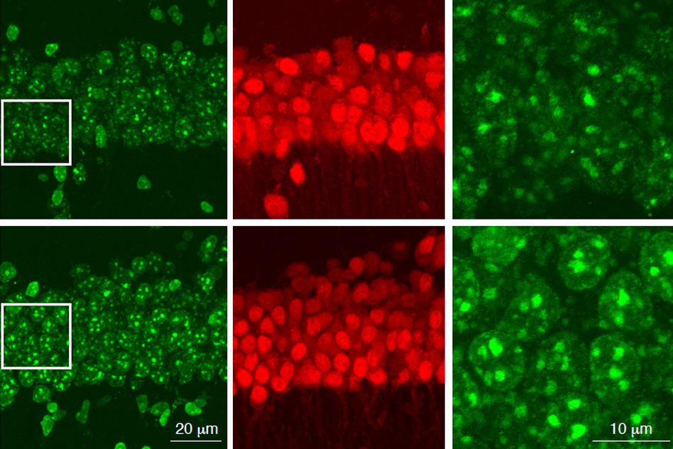
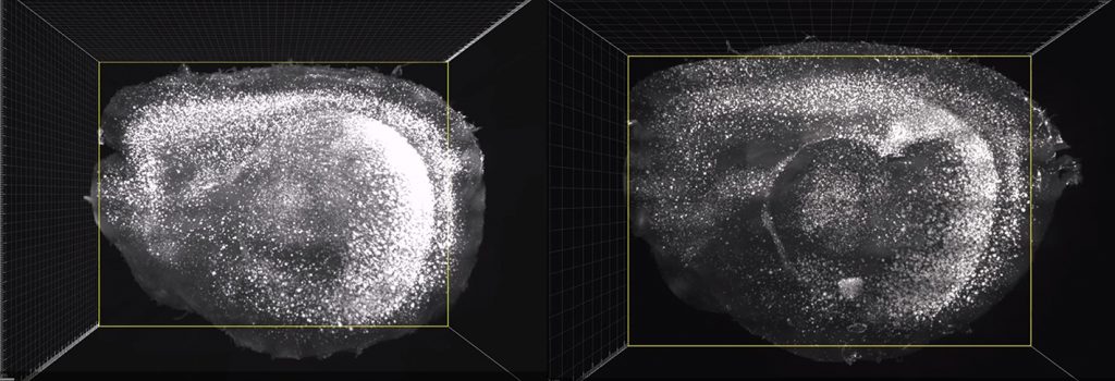
To investigate the connection between Alzheimer’s, the APOE4 variant and CAA, Blanchard, Tsai and co-authors coaxed human induced pluripotent stem cells to become the three types of cells that make up the BBB: brain endothelial cells, astrocytes and pericytes. Pericytes were modeled by mural cells that they tested extensively to ensure they exhibited pericyte-like properties and gene expression.

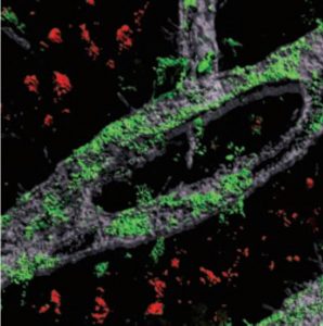
Grown for two weeks within a three-dimensional hydrogel scaffold, the BBB model cells assembled into vessels that exhibited natural BBB properties, including low permeability to molecules and expression of the same key genes, proteins and molecular pumps as natural BBBs. When immersed in culture media high in amyloid proteins, mimicking conditions in Alzheimer’s disease brains, the lab-grown BBB models exhibited the same kind of amyloid accumulation seen in human disease.

With a model BBB established, they then sought to test the difference APOE4 makes. They showed by several measures that APOE4-carrying BBB models accumulated more amyloid from culture media than those carrying APOE3, the more typical and healthy variant.

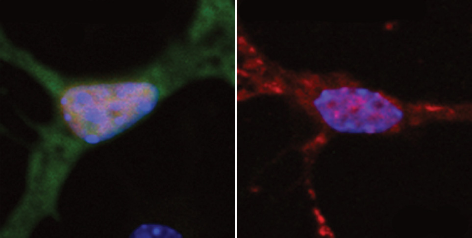
To pinpoint how APOE4 makes that difference, they engineered eight different versions covering all the possible combinations of the three cell types having either APOE3 or APOE4. When exposed these month-old models to amyloid-rich media, only versions with APOE4 pericyte-like mural cells showed excessive accumulation of amyloid proteins. Replacing APOE4 mural cells with APOE3-carrying ones reduced amyloid deposition. These results put blame for CAA-like pathology squarely on pericytes.

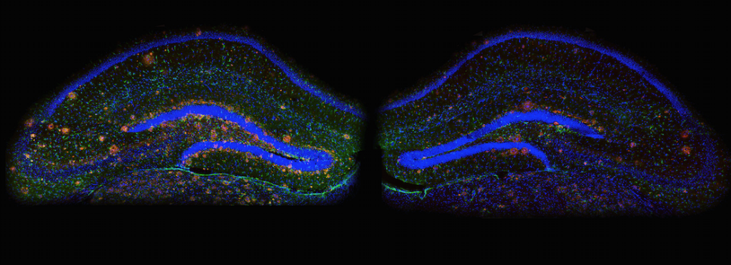
To further validate the clinical relevance of these findings, the team also looked at APOE expression in samples of human brain vasculature in the prefrontal cortex and the hippocampus, two regions crucially affected in Alzheimer’s disease. Consistent with the team’s lab BBB model, people with APOE4 showed higher expression of the gene in the vasculature, and specifically in pericytes, than people with APOE3.

“That is a salient point of this paper,” said Tsai, a founding member of MIT’s Aging Brain Initiative. “It’s really cool because it stresses the cell-type specific function of APOE.”

A pathway toward treatment?

The next step was to determine how APOE4 becomes so overexpressed by pericytes. The team therefore identified hundreds of transcription factors – proteins that determine how genes are expressed – that were regulated differently between APOE3 and APOE4 pericyte-like mural cells. Then they scoured that list to see which factors specifically impact APOE expression. A set of factors that were upregulated in APOE4 cells stood out: ones that were part of the calcineurin/NFAT pathway. They observed similar upregulation of the pathway in pericytes from human hippocampus samples.

As part of their investigation of whether elevated signaling activity of this pathway caused increased amyloid deposition and CAA, they tested cyclosporine A and FK506 because they tamp pathway activity down. They found that the drugs reduced APOE expression in their pericyte-like mural cells and therefore APOE4-mediated amyloid deposits in the BBB models. They also tested the drugs in APOE4-carrying mice and saw that the medicines reduced APOE expression and amyloid buildup.

Blanchard and Tsai noted that the drugs can have significant side effects, so their findings might not suggest using exactly those drugs to address CAA in patients.

“Instead it points toward the value of understanding the mechanism,” Blanchard said. “It allows one to design a small molecule screen to find more potent drugs that have less off-target effects.”

In addition to Blanchard and Tsai, the paper’s other authors are Michael Bula, Jose Davila-Velderrain, Leyla Akay, Lena Zhu, Alexander Frank, Matheus Victor, Julia Maeve Bonner, Hansruedi Mathys, Yuan-Ta Lin, Tak Ko, David Bennett, Hugh Cam, and Manolis Kellis.

The Robert A. and Renee E. Belfer Family Foundation, the Cure Alzheimer’s Fund, The National Institutes of Health, the Glenn Foundation for Medical Research and the American Federation for Aging Research funded the research.

A six panel grid showing neurons stained in red or green

Study finds that aging neurons accumulate DNA damage

MIT neuroscientists have discovered that an enzyme called HDAC1 is critical for repairing age-related DNA damage to genes involved in memory and other cognitive functions. This enzyme is often diminished in both Alzheimer’s patients and normally aging adults. In a study of mice, the researchers showed that when HDAC1 is lost, a specific type of DNA damage builds up as the mice age. They also showed that they could reverse this damage and improve cognitive function with a drug that activates HDAC1.

The study suggests that restoring HDAC1 could have positive benefits for both Alzheimer’s patients and people who suffer from age-related cognitive decline, the researchers say.

“It seems that HDAC1 is really an anti-aging molecule,” says Li-Huei Tsai, the director of MIT’s Picower Institute for Learning and Memory and the senior author of the study. “I think this is a very broadly applicable basic biology finding, because nearly all of the human neurodegenerative diseases only happen during aging. I would speculate that activating HDAC1 is beneficial in many conditions.”

Picower Institute research scientist Ping-Chieh Pao is the lead author of the study, which appears today in Nature Communications.

DNA repair and aging

There are several members of the HDAC family of enzymes, and their primary function is to modify histones — proteins around which DNA is spooled. These modifications control gene expression by blocking genes in certain stretches of DNA from being copied into RNA.

In 2013, Tsai’s lab published two papers that linked HDAC1 to DNA repair in neurons. In the current paper, the researchers explored what happens when HDAC1-mediated repair fails to occur. To do that, they engineered mice in which they could knock out HDAC1 specifically in neurons and another type of brain cells called astrocytes.

For the first several months of the mice’s lives, there were no discernable differences in their DNA damage levels or behavior, compared to normal mice. However, as the mice aged, differences became more apparent. DNA damage began to accumulate in the HDAC1-deficient mice, and they also lost some of their ability to modulate synaptic plasticity — changes in the strength of the connections between neurons. The older mice lacking HCAC1 also showed impairments in tests of memory and spatial navigation.

The researchers found that HDAC1 loss led to a specific type of DNA damage called 8-oxo-guanine lesions, which are a signature of oxidative DNA damage. Studies of Alzheimer’s patients have also shown high levels of this type of DNA damage, which is often caused by accumulation of harmful metabolic byproducts. The brain’s ability to clear these byproducts often diminishes with age.

An enzyme called OGG1 is responsible for repairing this type of oxidative DNA damage, and the researchers found that HDAC1 is needed to activate OGG1. When HDAC1 is missing, OGG1 fails to turn on and DNA damage goes unrepaired. Many of the genes that the researchers found to be most susceptible to this type of damage encode ion channels, which are critical for the function of synapses.

Targeting neurodegeneration

Several years ago, Tsai and Stephen Haggarty of Harvard Medical School, who is also an author of the new study, screened libraries of small molecules in search of potential drug compounds that activate or inhibit members of the HDAC family. In the new paper, Tsai and Pao used one of these drugs, called exifone, to see if they could reverse the age-related DNA damage they saw in mice lacking HDAC1.

The researchers used exifone to treat two different mouse models of Alzheimer’s, as well as healthy older mice. In all cases, they found that the drug reduced the levels of oxidative DNA damage in the brain and improved the mice’s cognitive functions, including memory.

Exifone was approved in the 1980s in Europe to treat dementia but was later taken off the market because it caused liver damage in some patients. Tsai says she is optimistic that other, safer HDAC1-activating drugs could be worth pursuing as potential treatments for both age-related cognitive decline and Alzheimer’s disease.

“This study really positions HDAC1 as a potential new drug target for age-related phenotypes, as well as neurodegeneration-associated pathology and phenotypes,” she says.

Tsai’s lab is now exploring whether DNA damage and HDAC1 also play a role in the formation of Tau tangles — misfolded proteins in the brain that are a signature of Alzheimer’s and other neurodegenerative diseases.

The research was funded by the National Institute on Aging, the National Institute of Neurological Disorders and Stroke, and a Glenn Award for Research in Biological Mechanisms of Aging.

–From MIT News

In mouse brains stained for the presence of amyloid, much less is visible in the cortex of a mouse treated with sensory gamma stimulation (right) than in a mouse left untreated (left).

In Cell: Brain wave stimulation may improve Alzheimer’s symptoms

By exposing mice to a unique combination of light and sound, MIT neuroscientists have shown that they can improve cognitive and memory impairments similar to those seen in Alzheimer’s patients.

This noninvasive treatment, which works by inducing brain waves known as gamma oscillations, also greatly reduced the number of amyloid plaques found in the brains of these mice. Plaques were cleared in large swaths of the brain, including areas critical for cognitive functions such as learning and memory.

“When we combine visual and auditory stimulation for a week, we see the engagement of the prefrontal cortex and a very dramatic reduction of amyloid,” says Li-Huei Tsai, director of MIT’s Picower Institute for Learning and Memory and the senior author of the study.

Further study will be needed, she says, to determine if this type of treatment will work in human patients. The researchers have already performed some preliminary safety tests of this type of stimulation in healthy human subjects.

MIT graduate student Anthony Martorell and Georgia Tech graduate student Abigail Paulson are the lead authors of the study, which appears in the March 14 issue of Cell.

Continue reading…

Review: DNA Damage and Its Links to Neurodegeneration

The integrity of our genetic material is under constant attack from numerous endogenous and exogenous agents. The consequences of a defective DNA damage response are well studied in proliferating cells, especially with regards to the development of cancer, yet its precise roles in the nervous system are relatively poorly understood. Here we attempt to provide a comprehensive overview of the consequences of genomic instability in the nervous system. We highlight the neuropathology of congenital syndromes that result from mutations in DNA repair factors and underscore the importance of the DNA damage response in neural development. In addition, we describe the findings of recent studies, which reveal that a robust DNA damage response is also intimately connected to aging and the manifestation of age-related neurodegenerative disorders such as Alzheimer’s disease and amyotrophic lateral sclerosis.

Video Abstract

read more →

Neuroscientists find that limiting a certain protein in the brain reverses Alzheimer’s symptoms in mice

Limiting a certain protein in the brain reverses Alzheimer’s symptoms in mice, report neuroscientists at MIT’s Picower Intitute for Learning and Memory.

Researchers found that the overproduction of the protein known as p25 may be the culprit behind the sticky protein-fragment clusters that build up in the brains of Alzheimer’s patients. The work, which was published in the April 10 issue of Cell, could provide a new drug target for the treatment of the disease that affects more than five million Americans, says Li-Huei Tsai, director of MIT’s Picower Institute for Learning and Memory and senior author of the paper.

read more →

Drug Tweaks Epigenome to Erase Fear Memories

A hurricane, a car accident, a roadside bomb, a rape — extreme stress is more common than you might think, with an estimated 50 to 60 percent of Americans experiencing it at some point in their lives. About 8 percent of that group will be diagnosed with post-traumatic stress disorder, or PTSD. They will have flashbacks and nightmares. They will feel amped up, with nerves on a permanent state of high alert. They won’t be able to forget.

read more →

Li-Huei Tsai

Science In Mind

MIT researchers find a drug that helps erase traumatic memories in mice.

For years, neuroscientist Li-Huei Tsai has been unraveling the brain circuits that underlie memory, searching for approaches that might be helpful in treating Alzheimer’s disease. In 2007, the Massachusetts Institute of Technology scientist identified an experimental drug that could restore lost memories in mice. Lately, she has been wondering whether that kind of drug might be useful to help people forget traumatic events that cause fear and anxiety.

In a study published Thursday in the journal Cell, Tsai and colleagues used a single dose of the drug, called an HDAC inhibitor, to help mice extinguish a fearful memory of a traumatic event that took place in the distant past.

Read more →

By Carolyn Y. Johnson / Globe Staff

Erasing Traumatic Memories

Erasing traumatic memories

New study identifies drug that could improve treatment of posttraumatic stress disorder.

Nearly 8 million Americans suffer from posttraumatic stress disorder (PTSD), a condition marked by severe anxiety stemming from a traumatic event such as a battle or violent attack.

Many patients undergo psychotherapy designed to help them re-experience their traumatic memory in a safe environment so as to help them make sense of the events and overcome their fear. However, such memories can be so entrenched that this therapy doesn’t always work, especially when the traumatic event occurred many years earlier.

MIT neuroscientists have now shown that they can extinguish well-established traumatic memories in mice by giving them a type of drug called an HDAC2 inhibitor, which makes the brain’s memories more malleable, under the right conditions. Giving this type of drug to human patients receiving psychotherapy may be much more effective than psychotherapy alone, says Li-Huei Tsai, director of MIT’s Picower Institute for Learning and Memory.

Illustration: Christine Daniloff/MIT

Read more →

DNA damage may cause ALS

New study finds link between neurons’ inability to repair DNA and neurodegeneration.

Amyotrophic lateral sclerosis (ALS) — also known as Lou Gehrig’s disease — is a neurodegenerative disease that destroys the neurons that control muscle movement. There is no cure for ALS, which kills most patients within three to five years of the onset of symptoms, and about 5,600 new cases are diagnosed in the United States each year.

MIT neuroscientists have found new evidence that suggests that a failure to repair damaged DNA could underlie not only ALS, but also other neurodegenerative disorders such as Alzheimer’s disease. These findings imply that drugs that bolster neurons’ DNA-repair capacity could help ALS patients, says Li-Huei Tsai, director of MIT’s Picower Institute for Learning and Memory and senior author of a paper describing the ALS findings in the Sept. 15 issue of Nature Neuroscience.

Learn more →