Category: In The News

ON a black background the M-shaped coronal cross section of a mouse brain featurse patches of magenta and teal dots

With fractured genomes, Alzheimer’s neurons call for help

Study indicates that ailing neurons may instigate an inflammatory response from the brain’s microglia immune cells

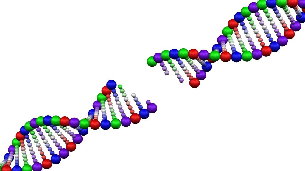
A new study by researchers in The Picower Institute for Learning and Memory at MIT provides evidence from both mouse models and postmortem human tissue of a direct link between two problems that emerge in Alzheimer’s disease: a buildup of double-stranded breaks (DSBs) in the DNA of neurons and the inflammatory behavior of microglia, the brain’s immune cells.

A key new finding is that neurons actively trigger an inflammatory response to their genomic damage. Neurons have not been known to signal the brain’s immune system in Alzheimer’s disease, said study lead author Gwyneth Welch, a former MIT Brain and Cognitive Sciences graduate student in the lab of senior author Li-Huei Tsai.

“This is a novel concept in neuroscience: the idea that neurons can be activating inflammatory activity in response to DNA damage,” Welch said. “The general idea was that neurons have a more passive relationship with microglia regarding age-associated neuroinflammation.”

Instead, what Welch, Tsai and co-authors report in Science Advances is that neurons coping with mounting DSBs go through stages of first trying to fix their fractured DNA and then, when it apparently fails, sending out via molecular signals to microglia, which responded by taking on a more inflammatory state. In experiments where the scientists interrupted the immune signaling, they prevented microglia from entering that state and degrading neural circuit connections, or synapses.

Members of Tsai’s lab have been studying DSBs in the context of Alzheimer’s for more than a decade. Tsai said the new findings add to the emerging understanding of the role they play in Alzheimer’s.

“We have a longstanding interest in understanding DNA breaks in neurons,” said Tsai, Picower Professor of Neuroscience and a founder of MIT’s Aging Brain Initiative. “We previously showed that DNA double stranded breaks are necessary for the induction of activity-regulated gene expression in neurons but we also observed profound DNA damage in neurons in the early stages of neurodegeneration.

“We now know that DNA-damaged neurons exhibit senescent phenotypes and play an active role in eliciting an immune response from microglia and perhaps astrocytes,” Tsai said. “This is mediated by the activation of the NFkappaB transcription factor. Moreover, we identified two cytokines secreted by damaged neurons to recruit microglia and elicit microglial response.  Importantly, we show that inhibition of NFkappaB rescued synaptic loss in neurodegeneration, further elucidating of impact of neuroimmune response on synaptic integrity and cognitive function.”

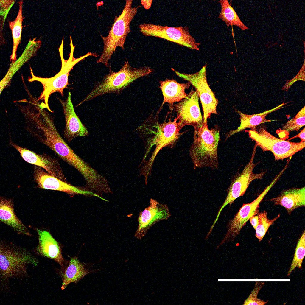
In her thesis research Welch used the lab’s “CK-p25” mouse model of Alzheimer’s, in which disease pathology can be induced, She observed a timeline in which neurons with DSBs appeared within a week, peaked in number after two weeks and then tapered off, becoming notably reduced by six weeks. Meanwhile, those neurons also lost their ability to express a standard marker of neuronal identity. Welch realized there seemed to be stages to the process of coping with DSBs. First neurons have few DSBs and strong identity (baseline), then high DSBs with no loss of identity (stage 1), then high DSBs and a loss of neuronal identity (stage 2).

Transcripts tell the tale

To understand what cells were doing differently in each stage, Welch and the team used multiple “transcriptomics” technologies, which tracks differences in gene expression. Her analyses revealed that neuronal identity genes were most strongly expressed at baseline, DNA repair genes were strong during stage 1, and immune signaling genes were particularly prominent during stage 2.

Among the immune signaling genes were ones governed by the master transcription regulator NFkappaB. These included the cytokines Ccl2 and Cxcl10.

To see if these changes were due specifically to DSBs, Welch treated neurons in the absence of any induced pathology with a chemical called etoposide that causes DSBs. Similar gene expression patterns evident in the induced mice were recapitulated in the ones treated with etoposide. And when Welch also looked at gene expression in the brains of people with DSBs and with Alzheimer’s she also found many significant overlaps.

“We found that stage 1 and stage 2 gene signatures were active in human DSB-bearing neurons,” she and her co-authors wrote. “This neuronal immune signature was further amplified in the context of AD pathology, suggesting that it may serve a functional role in disease-associated neuroinflammation.”

A big role for microglia

Having established that DSB-afflicted neurons employ NFkappaB to send out immune signals such as Ccl2 and Cxcl10, Welch and the team then asked what the effect was. Given that the lab in 2017 had characterized a late-stage inflammatory response on the part of microglia in Alzheimer’s, they hypothesized that neurons might be responsible.

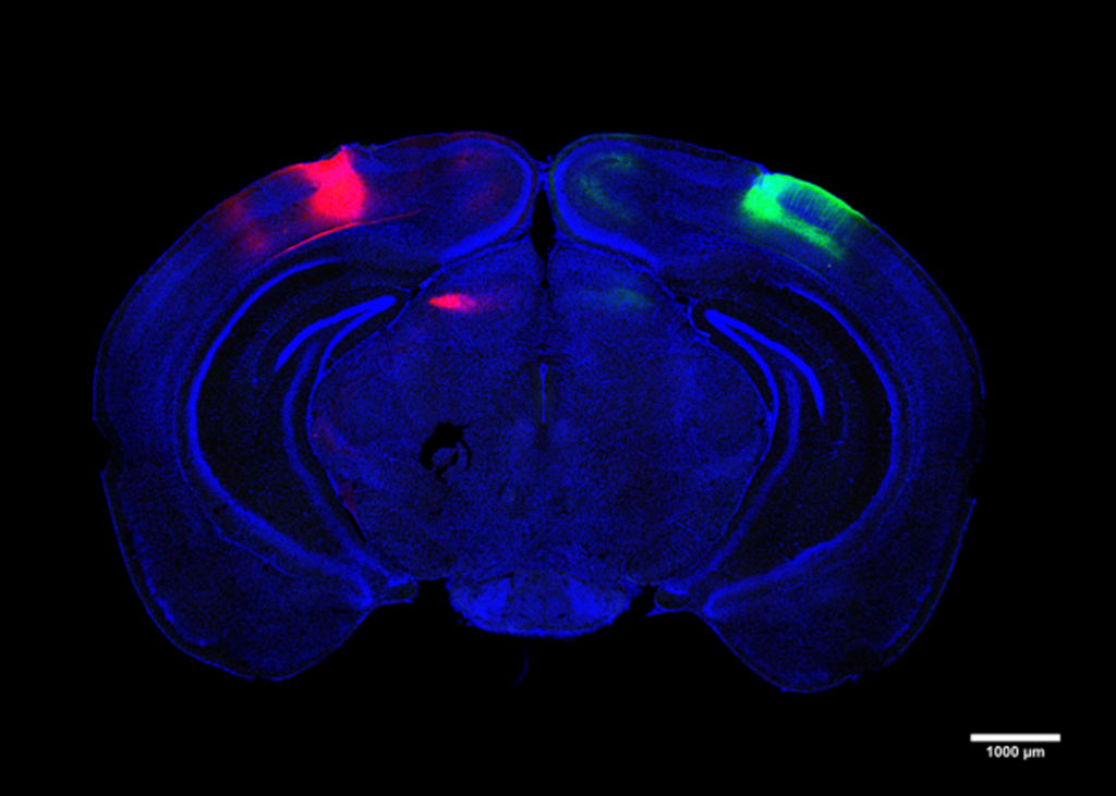
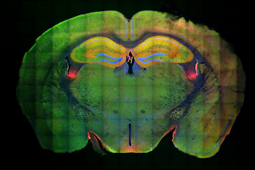
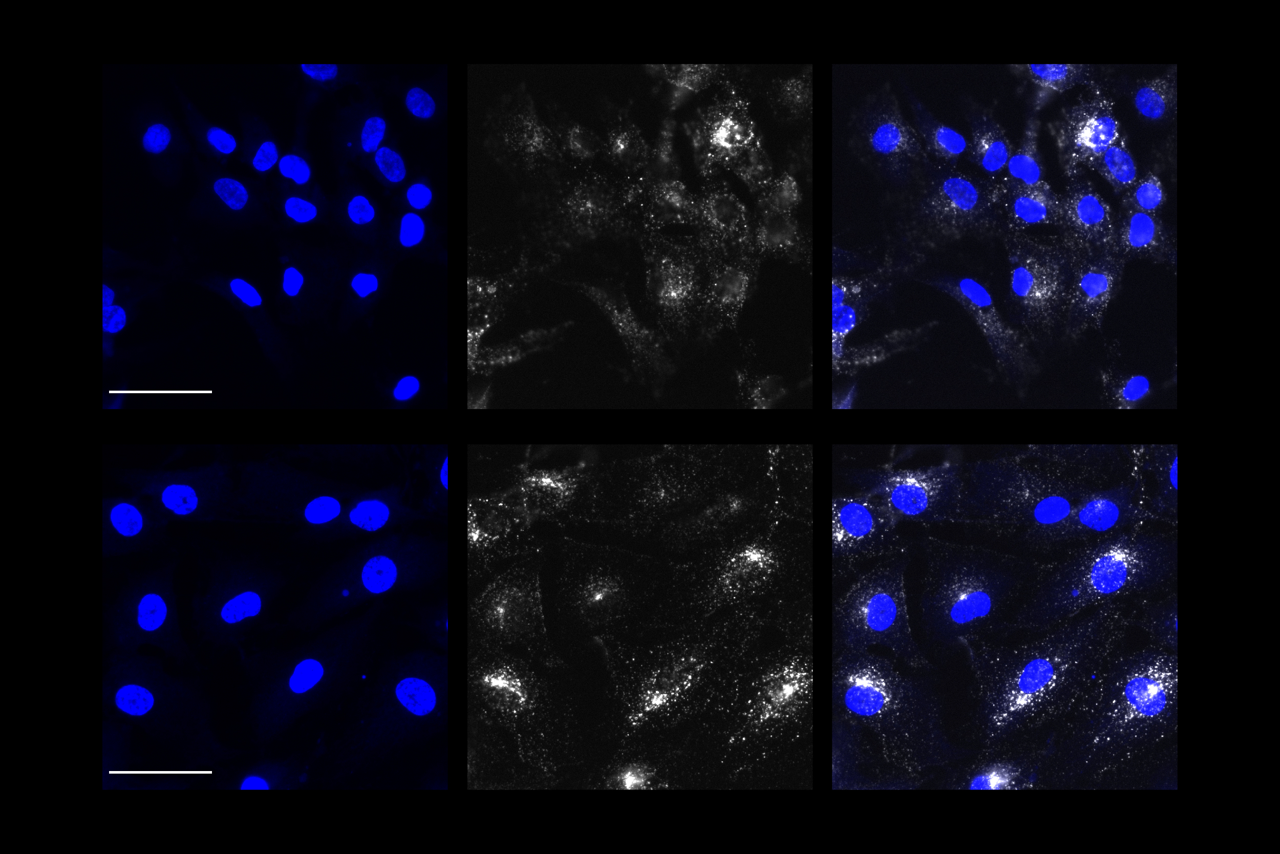
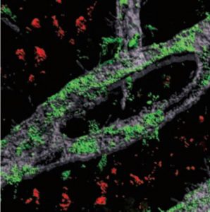
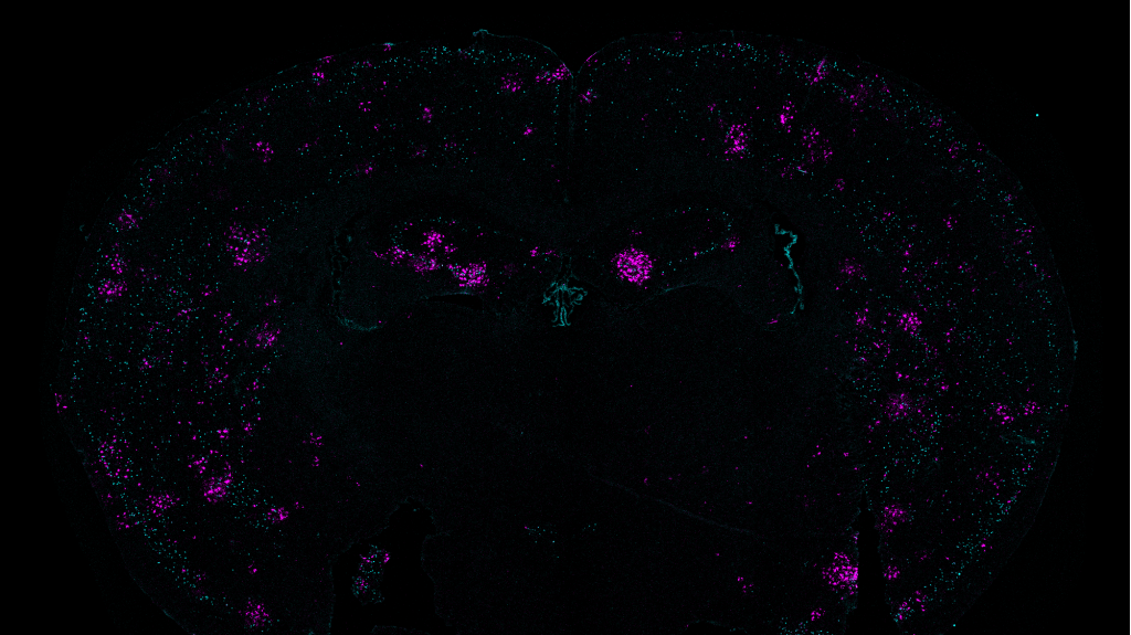
For this analysis Welch used spatial transcriptomics. She divided up both uninduced and induced mouse brains into many areas, and rated each area based on the strength of their DSB signal. Then she analyzed gene transcription in each area and found that locations with high DSBs also had many more microglia in an inflammatory state than locations with low DSBs. They were also able to directly image this relationship, yielding the observation that inflammatory microglia (evidenced by abnormally large cell bodies) were co-located with high-DSB neurons.

To further test the hypothesis, they disrupted NFkappaB regulated transcription in neurons by interfering with a key molecular cog in that machinery called p65. That step resulted in reduced proliferation of microglia and reduced microglia cell body size. It also induced beneficial changes in microglia gene expression, making them more consistent with their normal “homeostatic” state.

In other experiments they found that etoposide-treated neurons expressed Cxcl10 and Ccl2 but that disrupting NFkappaB reduced that expression. They also saw that depleting the two molecules from the brains also prevented microglia from becoming reactive.

And looking back at the neurons, they saw that while knocking down NFkappaB activity didn’t prevent them from dying, it did preserve circuit connections, or synapses, on the neurons that remained alive. That’s important because those circuit connections underlie brain function and microglia are known to prune those.

Because NFkappaB is known to help prevent cell death (which may be why knocking it down didn’t prevent neurons from dying), Welch said knocking it down is not likely to be a therapeutic strategy.

“Its more a proof of principle that that if you turn off a major switch for inflammation, that will change how microglia and neurons interact,” she said. “If your goal is to target inflammatory pathways, focusing on specific signaling molecules might be the more precise way to intervene.”

In addition to Welch and Tsai, the paper’s other authors are Carles Boix, Eloi Schmauch, Jose Davila-Velderrain, Matheus Victor, Vishnu Dileep, P. Lorenzo Bozzelli, Qiao Su, Jemmie D. Cheng, Audrey Lee, Noelle S. Leary, Andreas R. Pfenning, and Manolis Kellis.

The National Institutes of Health, CureAlz, the Glenn Foundation, and the JPB Foundation provided funding for the research.

A blue-hued cross-secction of a mouse;s brain highlights the visual cortex in red on the upper left and green on the upperright.

When Alzheimer’s degrades cells that cross hemispheres, visual memory suffers

New research reveals cells that span brain hemispheres to coordinate activity in visual processing centers and shows that Alzheimer’s degrades their structure and therefore their function

A new MIT study finds that Alzheimer’s disease disrupts at least one form of visual memory by degrading a newly identified circuit that connects the vision processing centers of each brain hemisphere.

The results of the study, published in Neuron by a research team based at The Picower Institute for Learning and Memory, come from experiments in mice, but provide a physiological and mechanistic basis for prior observations in human patients: the degree of diminished brain rhythm synchrony between counterpart regions in each hemisphere correlates with the clinical severity of dementia.

“We demonstrate that there is a functional circuit that can explain this phenomenon,” said lead author Chinnakkaruppan Adaikkan, a former Picower Institute postdoc who is now an assistant professor in the Centre for Brain Research at the Indian Institute of Science (IISc) in Bangalore. “In a way we uncovered a fundamental biology that was not known before.”

Specifically, Adaikkan’s work identified neurons that connect the primary visual cortex (V1) of each hemisphere and showed that when the cells are disrupted, either by genetic alterations that model Alzheimer’s disease or by direct laboratory perturbations, brain rhythm synchrony becomes reduced and mice become significantly less able to notice when a new pattern appeared on a wall in their enclosures. Such recognition of novelty, which requires visual memory of what was there the prior day, is an ability commonly disrupted in Alzheimer’s.

“This study demonstrates the propagation of gamma rhythm synchrony across the brain hemispheres via the cross hemispheric connectivity,” said study senior author Li-Huei Tsai, Picower Professor and director of The Picower Institute and MIT’s Aging Brain Initiative. “It also demonstrates that the disruption of this circuit in AD mouse models is associated with specific behavioral deficits.”

Cross-hemispheric cells

In the study, Adaikkan, Tsai, Thomas McHugh and co-authors discovered and traced V1 neurons that extended their axons all the way through the corpus callosum, which connects the brain’s hemispheres, to cells in the V1 on the brain’s other side. There, they found, the cross-hemispheric (CH) neurons forged connections, or synapses, with target cells, providing them with “excitatory” stimulation to drive their activity. Adaikkan also found that CH neurons were much more likely to be activated by a novelty discrimination task than V1 neurons in general or neurons in other regions heavily involved in memory such as the hippocampus or the prefrontal cortex.

Curious about how this might differ in Alzheimer’s disease, the team looked at the activity of the cells in two different Alzheimer’s mouse models. The found that CH cell activity was significantly lessened amid the disease. Unsurprisingly, Alzheimer’s mice fared much poorer in novelty discrimination tasks.

The team examined the CH cells closely and found that they gather incoming input from a variety of other cells within their V1 and other regions in their hemisphere that process visual information. When they compared the incoming connections of healthy CH neurons to those in CH cells afflicted with Alzheimer’s, they found that cells in the disease condition had significantly less infrastructure for hosting incoming connections (measured in terms of synapse-hosting spines protruding from the vine-like dendrites that sprawl out of the cell body).

Given the observations correlating reduced brain rhythm synchrony and memory performance in Alzheimer’s, the team wondered if that occurred in the mice, too. To find out, they custom-designed electrodes to measure rhythmic activity simultaneously in all cortical layers of each hemisphere’s V1. They observed that cross-hemispheric synchrony increased notably between the V1s when mice engaged in novelty discrimination but that the synchrony, both at high “gamma” and lower “theta” frequency rhythms, was significantly lower in the Alzheimer’s mice than it was in healthy mice.

Adaikkan’s evidence at that point was strong, but still only suggestive, that CH neurons provided the means by which the V1 regions on each side of the brain could coordinate to enable novelty discrimination, and that this ability became undermined by Alzheimer’s degradation of the CH cells’ connectivity. To more directly determine whether the CH circuit played such a causal, consequential role, the team directly intervened to disrupt them, testing what effect targeted perturbations had.

They found that chemically inhibiting CH cells disrupted rhythm synchrony between V1s, mirroring measures made in Alzheimer’s model mice. Moreover, disrupting CH activity undermined novelty discrimination ability. To further test whether it was the cells’ cross-hemispheric nature that mattered specifically, they engineered CH cells to be controllable with flashes of light (a technology called “optogenetics”). When they shined the light on the connections, they forged in the other hemisphere to inhibit those, they found that doing so again compromised visual discrimination ability.

All together, the study results show that CH cells in V1 connect with neurons in the counterpart area of the opposite hemisphere to synchronize neural activity needed for properly recognizing novelty, but that Alzheimer’s disease damages their ability to do that job.

Adaikkan said he is curious to now look at other potential cross-hemispheric connections and how they may be affected in Alzheimer’s disease, too. He said he also wants to study what happens to synchrony at other rhythm frequencies.

In addition to Adaikkan and Tsai, the study’s other authors are Jun Wang, Karim Abdelall, Steven Middleton, Lorenzo Bozzelli, and Ian Wickersham.

The JPB Foundation, The National Institutes of Health, and the Robert A. and Renee E. Belfer Foundation provided funding for the study.

A swirling blue ball of long thin neurons is overlaid with long electrodes coming in from the bottom and the left.

How microglia contribute to Alzheimer’s disease

A breakdown of lipid metabolism in these brain cells promotes inflammation and interferes with neuron activity, a new study finds

One of the hallmarks of Alzheimer’s disease is a reduction in the firing of some neurons in the brain, which contributes to the cognitive decline that patients experience. A new study from MIT shows how a type of cells called microglia contribute to this slowdown of neuron activity.

The study found that microglia that express the APOE4 gene, one of the strongest genetic risk factors for Alzheimer’s disease, cannot metabolize lipids normally. This leads to a buildup of excess lipids that interferes with nearby neurons’ ability to communicate with each other.

“APOE4 is a major genetic risk factor, and many people carry it, so the hope is that by studying APOE4, that will also provide a bigger picture of the fundamental pathophysiology of Alzheimer’s disease and what fundamental cell processes that have to go wrong to result in Alzheimer’s disease,” says Li-Huei Tsai, director of MIT’s Picower Institute for Learning and Memory and the senior author of the study.

On a black background we see a nearly round mass of tangly blue neurons with flecks of green throughout
For their study researchers cultured spheroids of neurons. Here neurons are stained purple among more general cell nuclei (blue) and astrocyte cells (green).

The findings suggest that if researchers could find a way to restore normal lipid metabolism in microglia, that might help to treat some of the symptoms of the disease.

MIT postdoc Matheus Victor is the lead author of the paper, which appears today in Cell Stem Cell.

Lipid overload

About 14 percent of the population has the APOE4 variant, making it the most common genetic variant that has been linked to late-onset, nonfamilial Alzheimer’s disease. People who carry one copy of APOE4 have a threefold higher risk of developing Alzheimer’s, and people with two copies have a tenfold higher risk.

“If you look at this another way, if you look at the entire Alzheimer’s disease population, about 50 percent of them are APOE4 carriers. So, it’s a very significant risk, but we still don’t know why this APOE4 allele presents such a risk,” Tsai says.

The APOE gene also comes in two other forms, known as APOE2, which is considered protective against Alzheimer’s, and the most common form, APOE3, which is considered neutral. APOE3 and APOE4 differ by just one amino acid.

For several years, Tsai’s lab has been studying the effects of APOE4 on a variety of cell types in the brain. To do this, the researchers use induced pluripotent stem cells, derived from human donors, and engineer them to express a specific version of the APOE gene. These cells can then be stimulated to differentiate into brain cells, including neurons, microglia, and astrocytes.

In a 2018 study, they showed that APOE4 causes neurons to produce large quantities of amyloid beta peptide 42, an Alzheimer’s-linked molecule that causes the neurons to become hyperactive. That study found that APOE4 also affects the functions of microglia and astrocytes, leading to cholesterol accumulation, inflammation, and failure to clear amyloid beta peptides.

A 2021 follow-up showed that APOE4 astrocytes have dramatic impairments in their ability to process a variety of lipids, which leads to a buildup of molecules such as triglycerides, as well as cholesterol. In that paper, the researchers also showed that treating engineered yeast cells expressing APOE4 with choline, a dietary supplement that is a building block for phospholipids, could reverse many of the detrimental effects of APOE4.

In their new study, the researchers wanted to investigate how APOE4 affects interactions between microglia and neurons. Recent research has shown that microglia play an important role in modulating neuronal activity, including their ability to communicate within neural ensembles. Microglia also scavenge the brain looking for signs of damage or pathogens, and clear out debris.

The researchers found that APOE4 disrupts microglia’s ability to metabolize lipids and prevents them from removing lipids from their environment. This leads to a buildup of fatty molecules, especially cholesterol, in the environment. These fatty molecules bind to a specific type of potassium channel embedded in neuron cell membranes, which suppresses neuron firing.

“We know that in late stages of Alzheimer’s disease, there is reduced neuron excitability, so we may be mimicking that with this model,” Victor says.

The buildup of lipids in microglia can also lead to inflammation, the researchers found, and this type of inflammation is believed to contribute to the progression of Alzheimer’s disease.

Restoring function

The researchers also showed that they could reverse the effects of lipid overload by treating APOE4 microglia with a drug called Triacsin C, which interferes with the formation of lipid droplets. When APOE4 microglia were exposed to this drug, the researchers found that normal communication between neighboring neurons and microglia was restored.

“We can rescue the suppression of neuronal activity by APOE4 microglia, presumably through lipid homeostasis being restored, where now fatty acids are not accumulating extracellularly,” Victor says.

Triacsin C can be toxic to cells, so it would likely not be suitable to use as a drug to treat Alzheimer’s, but the researchers hope that other approaches to restore lipid homeostasis could help combat the disease. In Tsai’s 2021 APOE4 study, she showed that choline also helps to restore normal microglia activity.

“Lipid homeostasis is actually critical for a number of cell types across the Alzheimer’s disease brain, so it’s not singularly a microglia problem,” Victor says. “The question is, how do you restore lipid homeostasis across multiple cell types? It’s not an easy task, but we’re tackling that through choline, for example, which might be a really interesting angle.”

The researchers are now further studying how microglia transition from a healthy state to a “lipid-burdened,” inflammatory state, in hopes of discovering ways to block that transition. In previous studies in mice, they have shown that exposure to LED light flickering at a specific frequency can help to rejuvenate microglia, stimulating the cells to resume their normal functions.

The research was funded by the National Institutes of Health, the Howard Hughes Medical Institute Hanna H. Gray Postdoctoral Fellowship, The Robert A. and Renee E. Belfer Family Foundation, Carol and Eugene Ludwig Family Foundation, the Cure Alzheimer’s Fund, The JPB Foundation, Joseph P. DiSabato and Nancy E. Sakamoto, Donald A. and Glenda G. Mattes, Lester A. Gimpelson, the Halis Family Foundation, the Dolby family, David Emmes, and Alan and Susan Patricof.

–From MIT News
Three bright yellow-green celll cultures on black background

In Down syndrome cells, genome-wide disruptions mimic a senescence-like state

Extra chromosome alters chromosomal conformation and DNA accessibility across the whole genome in neural progenitor cells, disrupting gene transcription and cell functions much like in cellular aging

In Down syndrome, the third copy of chromosome 21 causes a reorganization of the 3D configuration of the entire genome in a key cell type of the developing brain, a new study shows. The resulting disruption of gene transcription and cell function are so similar to those seen in cellular aging, or senescence, that the scientists leading the study found they could use anti-senescence drugs to correct them in cell cultures.

The study published in Cell Stem Cell therefore establishes senescence as a potentially targetable mechanism for future treatment of Down syndrome, said Hiruy Meharena, a new assistant professor at the University of California San Diego who led the work as a Senior Alana Fellow in the Alana Down Syndrome Center at MIT.

“There is a cell-type specific genome-wide disruption that is independent of the gene dosage response,” Meharena said. “It’s a very similar phenomenon to what’s observed in senescence. This suggests that excessive senescence in the developing brain induced by the third copy of chromosome 21 could be a key reason for the neurodevelopmental abnormalities seen in Down syndrome.”

The study’s finding that neural progenitor cells (NPCs), which develop into major cells in the brain including neurons, have a senescent character is remarkable and novel, said senior author Li-Huei Tsai, but it is substantiated by the team’s extensive work to elucidate the underlying mechanism of the effects of abnormal chromosome number, or aneupoloidy, within the nucleus of the cells.

“This study illustrates the importance of asking fundamental questions about the underlying mechanisms of neurological disorders,” said Tsai, Picower Professor of Neuroscience, director of the Alana Center, and of The Picower Institute for Learning and Memory at MIT. “We didn’t begin this work expecting to see senescence as a translationally relevant feature of Down syndrome, but the data emerged from asking how the presence of an extra chromosome affects the architecture of all of a cell’s chromosomes during development.”

Genomewide changes

Meharena and co-authors spent years measuring distinctions between human cell cultures that differed only by whether they had a third copy of chromosome 21. Stem cells derived from volunteers were cultured to turn into NPCs. In both the stem cells and the NPCs, the team examined 3D chromosome architecture, several metrics of DNA structure and interaction, gene accessibility and transcription, and gene expression. They also looked at the consequences of the gene expression differences on important functions of these developmental cells, such as how well they proliferated and migrated in 3D brain tissue cultures. Stem cells were not particularly different, but NPCs were substantially affected by the third copy of chromosome 21.

Li-Huei Tsai stands in a lab and points to an image of a cell on a computer screen. Hiruy Meharena sits at the desk with his hand on the mouse and looks on.
Li-Huei Tsai and Hiruy Meharena consult about images produced during the research in this 2019 photo

Overall, the picture that emerged in NPCs was that the presence of a third copy causes all the other chromosomes to squish inward, not unlike when people in a crowded elevator must narrow their stance when one more person squeezes in. The main effects of this “chromosomal introversion,” meticulously quantified in the study, are more genetic interactions within each chromosome and less interactions among them. These changes and differences in DNA conformation within the cell nucleus lead to changes in how genes are transcribed and therefore expressed, causing important differences in cell function that affect brain development.

Treated as senescence

For the first couple of years as these data emerged, Meharena said, the full significance of the genomic changes were not apparent, but then he read a paper showing very similar genomic rearrangement and transcriptional alterations in senescent cells.

After validating that the Down syndrome cells indeed bore such a similar signature of transcriptional differences, the team decided to test whether anti-senescence drugs could undo the effects. They tested a combination of two: dasatinib and quercetin. The medications improved not only gene accessibility and transcription, but also the migration and proliferation of cells.

That said, the drugs have very significant side effects—dasatinib is only given to cancer patients when other treatments have not done enough—so they are not appropriate for attempting to intervene in brain development amid Down syndrome, Meharena said. Instead an outcome of the study could be to inspire a search for medications that could have anti-senolytic effects with a safer profile.

A schematic shows the process of how a trisomy 21 cell experiences genomiwide chromosomal reconfiguration leading to a senescence-like response
A new study’s findings indicate that amid trisomy 21, neural progenitor cells experience a genome-wide chromosomal reorganization leading to a senescence-like response including altered chromatin states and gene transcription.

Senescence is a stress response of cells. At the same time, years of research by former MIT biology professor Angelika Amon, who co-directed the Alana Center with Tsai, has shown that aneuploidy is a source of considerable stress for cells. A question raised by the new findings, therefore, is whether the senescence-like character of Down syndrome NPCs is indeed the result of an aneuploidy induced stress and if so, exactly what that stress is.

Another implication of the findings is how excessive senescence among brain cells might affect people with Down syndrome later in life. The risk of Alzheimer’s disease is much higher at a substantially earlier age in the Down syndrome population than among people in general. In large part this is believed to be because a key Alzheimer’s risk gene, APP, is on chromosome 21, but the newly identified inclination for senescence may also accelerate Alzheimer’s development.

In addition to Meharena and Tsai, the paper’s other authors are Asaf Marco, Vishnu Dileep, Elana Lockshin, Grace Akatsu, James Mullahoo, Ashley Watson, Tak Ko, Lindsey Guerin, Fatema Abdurrob, Shruti Rengarajan, Malvina Papanastasiou and Jacob Jaffe.

The Alana Foundation, the LuMind Foundation, Burroughs Wellcome Fund, UNCF-Merck  and the National Institutes of Health funded the research.

A colorful cartoon of a DNA double helix with a gap through the middle

Memory making involves extensive DNA breaking

To quickly express genes needed for learning and memory, brain cells snap both strands of DNA in many more places and cell types than previously realized, a new study shows

The urgency to remember a dangerous experience requires the brain to make a series of potentially dangerous moves: Neurons and other brain cells snap open their DNA in numerous locations—more than previously realized, according to a new study—to provide quick access to genetic instructions for the mechanisms of memory storage.

The extent of these DNA double-strand breaks (DSBs) in multiple key brain regions is surprising and concerning, said study senior author Li-Huei Tsai, Picower Professor of Neuroscience at MIT and director of The Picower Institute for Learning and Memory, because while the breaks are routinely repaired, that process may become more flawed and fragile with age. Tsai’s lab has shown that lingering DSBs are associated with neurodegeneration and cognitive decline and that repair mechanisms can falter.

“We wanted to understand exactly how widespread and extensive this natural activity is in the brain upon memory formation because that can give us insight into how genomic instability could undermine brain health down the road,” said Tsai, who is also a professor in the Department of Brain and Cognitive Sciences and a leader of MIT’s Aging Brain Initiative. “Clearly memory formation is an urgent priority for healthy brain function but these new results showing that several types of brain cells break their DNA in so many places to quickly express genes is still striking.”

Tracking breaks

In 2015, Tsai’s lab provided the first demonstration that neuronal activity caused DSBs and that they induced rapid gene expression. But those findings, mostly made in lab preparations of neurons, did not capture the full extent of the activity in the context of memory formation in a behaving animal and did not investigate what happened in cells other than neurons.

IIn the new study published July 1 in PLOS ONE, lead author and former graduate student Ryan Stott and co-author and former research technician Oleg Kritsky sought to investigate the full landscape of DSB activity in learning and memory. To do so, they gave mice little electrical zaps to the feet when they entered a box, to condition a fear memory of that context. They then used several methods to assess DSBs and gene expression in the brains of the mice over the next half hour, particularly among a variety of cell types in the prefrontal cortex and hippocampus, two regions essential for the formation and storage of conditioned fear memories. They also made measurements in the brains of mice who did not experience the foot shock to establish a baseline of activity for comparison.

The creation of a fear memory doubled the number of DSBs among neurons in the hippocampus and the prefrontal cortex, affecting more than 300 genes in each region. Among 206 affected genes common to both regions, the researchers then looked at what those genes do. Many were associated with the function of the connections neurons make with each other, called synapses. This makes sense because learning arises when neurons change their connections (a phenomenon called “synaptic plasticity”) and memories are formed when groups of neurons connect together into ensembles called engrams.

“Many genes essential for neuronal function and memory formation, and significantly more of them than expected based on previous observations in cultured neurons…are potentially hotspots of DSB formation,” the authors wrote in the study.

In another analysis, the researchers confirmed through measurements of RNA that the increase in DSBs indeed correlated closely with increased transcription and expression of affected genes, including ones affecting synapse function, as quickly as 10-30 minutes after the foot shock exposure.

“Overall, we find transcriptional changes are more strongly associated with [DSBs] in the brain than anticipated,” they wrote. “Previously we observed 20 gene-associated [DSB] loci following stimulation of cultured neurons, while in the hippocampus and prefrontal cortex we see more than 100-150 gene associated [DSB] loci that are transcriptionally induced.”

Snapping with stress

In the analysis of gene expression, the neuroscientists looked at not only neurons but also non-neuronal brain cells, or glia, and found that they also showed changes in expression of hundreds of genes after fear conditioning. Glia called astrocytes are known to be involved in fear learning, for instance, and they showed significant DSB and gene expression changes after fear conditioning.

Among the most important functions of genes associated with fear conditioning-related DSBs in glia was the response to hormones. The researchers therefore looked to see which hormones might be particularly involved and discovered that it was glutocortocoids, which are secreted in response to stress. Sure enough, the study data showed that in glia, many of the DSBs that occurred following fear conditioning occurred at genomic sites related to glutocortocoid receptors. Further tests revealed that directly stimulating those hormone receptors could trigger the same DSBs that fear conditioning did and that blocking the receptors could prevent transcription of key genes after fear conditioning.

Tsai said the finding that glia are so deeply involved in establishing memories from fear conditioning is an important surprise of the new study.

“The ability of glia to mount a robust transcriptional response to glutocorticoids suggest that glia may have a much larger role to play in the response to stress and its impact on the brain during learning than previously appreciated,” she and her co-authors wrote.

Damage and danger?

More research will have to be done to prove that the DSBs required for forming and storing fear memories are a threat to later brain health, but the new study only adds to evidence that it may be the case, the authors said.

“Overall we have identified sites of DSBs at genes important for neuronal and glial functions, suggesting that impaired DNA repair of these recurrent DNA breaks which are generated as part of brain activity could result in genomic instability that contribute to aging and disease in the brain,” they wrote.

The National Institutes of Health, The Glenn Foundation for Medical Research and the JPB Foundation provided funding for the research.

Li-Huei Tsai is shown in profile as she stands smiling at a podium

Li-Huei Tsai elected to American Academy of Arts & Sciences

The American Academy of Arts & Sciences announced today that Li-Huei Tsai, Picower Professor of Neuroscience and Director of The Picower Institute for Learning & Memory, is among 252 luminaries elected to join its esteemed membership.

“We are honoring the excellence of these individuals, celebrating what they have achieved so far, and imagining what they will continue to accomplish,” said David Oxtoby, President of the American Academy, in the announcement. “The past year has been replete with evidence of how things can get worse; this is an opportunity to illuminate the importance of art, ideas, knowledge, and leadership that can make a better world.”

Tsai’s laboratory focuses on advancing understanding the molecular-, circuit- and systems-level mechanisms underlying neurodegenerative diseases such as Alzheimer’s. She has translated many of her lab’s fundamental insights into potential therapeutic approaches.

“I’m very honored to be elected to the academy and to be in the company of so many leading scholars and luminaries,” Tsai said. “The Academy’s mission of advancing the public good is also a philosophy that guides our work to understand and address neurodegenerative diseases such as Alzheimer’s disease. This recognition encourages us to continue our efforts with urgency and rigor.”

With her election, Tsai joins other Picower Institute faculty in the academy including Mark Bear, Emery N. Brown, Earl Miller, Mriganka Sur, Susumu Tonegawa and Matt Wilson. Also this year, four other MIT faculty members were elected to membership.

Study offers an explanation for why the APOE4 gene enhances Alzheimer’s risk

Gene variant disrupts lipid metabolism, but in experiments the effects were reversed by choline supplements

One of the most significant genetic risk factors for developing Alzheimer’s disease is a gene called APOE4, which is carried by almost half of all Alzheimer’s patients. A new study from MIT shows that this gene has widespread effects on brain cells’ ability to metabolize lipids and respond to stress.

In studies of human brain cells and yeast cells, the researchers found that the APOE4 gene significantly disrupts brain cells’ ability to carry out their normal functions. They also showed that treating these cells with extra choline, a widely available supplement that is considered safe for human use, could reverse many of these effects.

The researchers hope that their findings will lead to clinical studies of choline in people who carry the APOE4 gene, who make up about 14 percent of the overall population. Previous trials looking at choline’s effects on cognition showed mixed results, but those trials were not targeted specifically to people with the APOE4 gene.

“What we would really like to see is whether in the human population, in those APOE4 carriers, if they take choline supplements to a sufficient amount, whether that would delay or give them some protection against developing dementia or Alzheimer’s disease,” says Li-Huei Tsai, the director of MIT’s Picower Institute for Learning and Memory.

Tsai and the late Susan Lindquist, former director of MIT’s Whitehead Institute for Biomedical Research, are the senior authors of the study, which appears today in Science Translational Medicine. The paper’s three lead authors are former Whitehead and MIT postdocs Grzegorz Sienski and Priyanka Narayan, and current MIT postdoc Julia Maeve Bonner.

Lipid dysregulation

The human gene for APOE, or apolipoprotein E, comes in three versions. While APOE4 is linked to higher risk for Alzheimer’s, APOE2 is considered protective, and APOE3, the most common variant, is neutral.

APOE is known to be involved in lipid metabolism, but its role in the development of Alzheimer’s has been unclear, Tsai says. To try to learn more about this connection, the researchers created human induced pluripotent stem cells that carry either the APOE3 or APOE4 gene in an otherwise identical genetic background. They then stimulated these cells to differentiate into astrocytes, the brain cells that produce the most APOE.

APOE4 astrocytes showed dramatic changes in how they process lipids compared to APOE3. In APOE4 astrocytes, there was a significant buildup of neutral lipids and cholesterol. These astrocytes also accumulated droplets containing a type of lipids called triglycerides, and these triglycerides had many more unsaturated fatty acid chains than normal. These changes all disrupt the normal lipid balance inside the cells. The authors also noted APOE4-dependent lipid disruptions in another important brain cell, microglia.

“When lipid homeostasis is compromised, then a lot of very essential processes are affected, such as intracellular trafficking, vesicular trafficking, and endocytosis. A lot of the cells’ essential functions are compromised,” Tsai says.

“This balance is really important for cells to be able to perform normal functions like generate membranes and so on, but also to be able to absorb stress,” Bonner says. “We think that one of the things that’s happening is that these cells are less able to absorb stress because they’re already in this heightened lipid dysregulation state.”

The researchers also found that yeast cells engineered to express the human version of APOE4 showed many of the same defects. Using these cells, they performed a systematic genetic screen to determine the molecular basis of the defects seen in APOE4 cells. This screen showed that turning on a pathway that normally produces phospholipids, an essential component of cell membranes, can reverse some of the damage seen in APOE4 cells. This suggests that APOE4 somehow increases the requirement for phospholipid synthesis.

The researchers also found that growing APOE4 yeast cells on a very nutrient-rich growth medium helped them to survive better than APOE4 yeast cells grown on the typical growth medium. Further experiments revealed that the nutrient that helped APOE4 cells survive is choline, a building block that cells use to make phospholipids. The researchers then treated their human APOE4 astrocyte cells with choline to promote phospholipid synthesis, and found that it also reversed much of the damage they had seen in those cells, including the accumulation of cholesterol and lipid droplets.

Choline deficiency

The researchers have now begun studying a mouse model of Alzheimer’s that is also engineered to express the human APOE4 gene. They hope to investigate whether choline can help to reverse some of the symptoms of Alzheimer’s in these mice.

Choline is naturally found in foods such as eggs, meat, fish, and some beans and nuts. The minimum recommended intake of choline is 550 milligrams per day for men and 425 milligrams per day for women, but most people don’t consume that much, Tsai says. The new study offers preliminary evidence that people who carry the APOE4 gene may benefit from taking choline supplements, she says, although clinical trials are necessary to confirm that.

“What our results suggest is that if you are an APOE2 or APOE3 carrier, even you are somewhat choline deficient you can cope with it,” Tsai says. “But if you are an APOE4 carrier, then if you don’t take enough choline, then that will have a more dire consequences. The APOE4 carriers are more susceptible to choline deficiency.”

The research was funded by the EMBO Fellowship, the Helen Hay Whitney Foundation, the National Institutes of Health, the Damon Runyon Foundation, the Neurodegeneration Consortium, the Robert A. and Renee E. Belfer Foundation, the Howard Hughes Medical Institute, the Ludwig Family Foundation, and Kara and Stephen Ross.

–From MIT News

A cross section of a mouse brain stained in a rainbow of colors

Neuroscientists discover a molecular mechanism that allows memories to form

When the brain forms a memory of a new experience, neurons called engram cells encode the details of the memory and are later reactivated whenever we recall it. A new MIT study reveals that this process is controlled by large-scale remodeling of cells’ chromatin.

This remodeling, which allows specific genes involved in storing memories to become more active, takes place in multiple stages spread out over several days. Changes to the density and arrangement of chromatin, a highly compressed structure consisting of DNA and proteins called histones, can control how active specific genes are within a given cell.

“This paper is the first to really reveal this very mysterious process of how different waves of genes become activated, and what is the epigenetic mechanism underlying these different waves of gene expression,” says Li-Huei Tsai, the director of MIT’s Picower Institute for Learning and Memory and the senior author of the study.

Asaf Marco, an MIT postdoc, is the lead author of the paper, which appears today in Nature Neuroscience.


Above: The hippocampus is the large yellow structure near the top. Green indicates neurons that were activated in memory formation; red shows the neurons that were activated in memory recall; blue shows the DNA of the cells; and yellow shows neurons that were activated in both memory formation and recall, and are thus considered to be the engram neurons.


Epigenomic control

Engram cells are found in the hippocampus as well as other parts of the brain. Many recent studies have shown that these cells form networks that are associated with particular memories, and these networks are activated when that memory is recalled. However, the molecular mechanisms underlying the encoding and retrieval of these memories are not well-understood.

Neuroscientists know that in the very first stage of memory formation, genes known as immediate early genes are turned on in engram cells, but these genes soon return to normal activity levels. The MIT team wanted to explore what happens later in the process to coordinate the long-term storage of memories.

“The formation and preservation of memory is a very delicate and coordinated event that spreads over hours and days, and might be even months — we don’t know for sure,” Marco says. “During this process, there are a few waves of gene expression and protein synthesis that make the connections between the neurons stronger and faster.”

Tsai and Marco hypothesized that these waves could be controlled by epigenomic modifications, which are chemical alterations of chromatin that control whether a particular gene is accessible or not. Previous studies from Tsai’s lab have shown that when enzymes that make chromatin inaccessible are too active, they can interfere with the ability to form new memories.

To study epigenomic changes that occur in individual engram cells over time, the researchers used genetically engineered mice in which they can permanently tag engram cells in the hippocampus with a fluorescent protein when a memory is formed. These mice received a mild foot shock that they learned to associate with the cage in which they received the shock. When this memory forms, the hippocampal cells encoding the memory begin to produce a yellow fluorescent protein marker.

“Then we can track those neurons forever, and we can sort them out and ask what happens to them one hour after the foot shock, what happens five days after, and what happens when those neurons get reactivated during memory recall,” Marco says.

At the very first stage, right after a memory is formed, the researchers found that many regions of DNA undergo chromatin modifications. In these regions, the chromatin becomes looser, allowing the DNA to become more accessible. To the researchers’ surprise, nearly all of these regions were in stretches of DNA where no genes are found. These regions contain noncoding sequences called enhancers, which interact with genes to help turn them on. The researchers also found that in this early stage, the chromatin modifications did not have any effect on gene expression.

The researchers then analyzed engram cells five days after memory formation. They found that as memories were consolidated, or strengthened, over those five days, the 3D structure of the chromatin surrounding the enhancers changed, bringing the enhancers closer to their target genes. This still doesn’t turn on those genes, but it primes them to be expressed when the memory is recalled.

Next, the researchers placed some of the mice back into the chamber where they received the foot shock, reactivating the fearful memory. In engram cells from those mice, the researchers found that the primed enhancers interacted frequently with their target genes, leading to a surge in the expression of those genes.

Many of the genes turned on during memory recall are involved in promoting protein synthesis at the synapses, helping neurons strengthen their connections with other neurons. The researchers also found that the neurons’ dendrites — branched extensions that receive input from other neurons — developed more spines, offering further evidence that their connections were further strengthened.

Primed for expression

The study is the first to show that memory formation is driven by epigenomically priming enhancers to stimulate gene expression when a memory is recalled, Marco says.

“This is the first work that shows on the molecular level how the epigenome can be primed to gain accessibility. First, you make the enhancers more accessible, but the accessibility on its own is not sufficient. You need those regions to physically interact with the genes, which is the second phase,” he says. “We are now realizing that the 3D genome architecture plays a very significant role in orchestrating gene expression.”

The researchers did not explore how long these epigenomic modifications last, but Marco says he believes they may remain for weeks or even months. He now hopes to study how the chromatin of engram cells is affected by Alzheimer’s disease. Previous work from Tsai’s lab has shown that treating a mouse model of Alzheimer’s with an HDAC inhibitor, a drug that helps to reopen inaccessible chromatin, can help to restore lost memories.

The research was funded by the JBP Foundation and the Alzheimer’s Association.

–From MIT News

Long, thin,spiny cells stained yellow and blue appear above a black background

Alzheimer’s risk gene disrupts endocytosis, but another disease-linked gene could help

In a new study, a team of scientists based at The Picower Institute for Learning and Memory at MIT and the Whitehead Institute for Biomedical Research reveals evidence showing that the most prominent Alzheimer’s disease risk gene may disrupt a fundamental process in a key type of brain cell. Moreover, in a sign of how important it is to delve into the complex ways that genes intersect in disease, they found that increasing the expression of another Alzheimer’s-associated gene in those cells could help alleviate the problem.

About 25 percent of people have the APOE4 variant of the APOE gene, which puts them at substantially greater risk for Alzheimer’s disease than those with the more common APOE3 version. Scientists have been working for decades to understand why this is so. The new study in Cell Reports finds that in astrocytes, which are the most common non-neuron cell in the brain, the variant hampers the process of endocytosis, which is a major way that cells bring materials in from outside. That functional deficit could undermine several of the vital roles that astrocytes play in the brain, the researchers noted, including how they facilitate communication among neurons or maintain the blood-brain barrier, which stringently filters what circulates into or out of the brain.

“We have identified that APOE4 imposes an endocytosis deficiency in astrocytes,” said Priyanka Narayan, a researcher at the National Institutes of Health who co-led the work while a postdoc in the labs of the late Susan Lindquist, member of the Whitehead Institute, and of Li-Huei Tsai, Picower Professor of Neuroscience and the study’s corresponding author. “This effect could have a number of downstream consequences such as impaired communication with other cell types, poor clearance of extracellular material, or poor maintenance of metabolic homeostasis.”


Above: Astrocytes, such as these cells derived from induced pluripotent stem cells, are critical for brain function.


The research began in the lab of Lindquist, who was also a Professor of Biology at MIT. Lindquist and Tsai, were close collaborators. After Lindquist died, the research team completed the work in the Tsai lab at MIT. The study’s co-lead author is Grzegorz Sienski of the Whitehead Institute.

As part of their work, the team also found that in APOE4-carrying astrocytes increasing expression of an Alzheimer’s associated gene called PICALM reversed the endocytosis defects.

“Both APOE and PICALM are Alzheimer’s risk genes,” said Tsai, a founding director of MIT’s Aging Brain Initiative. “It is really interesting that the two genes converge on endocytosis. This indicates that faulty endocytosis plays a key role in the etiology of Alzheimer’s.”

Reduction and rescue

For at least a decade, studies have suggested connections among Alzheimer’s, APOE4 and errant endocytosis, but have not pinpointed specific mechanisms. The team sought them out—and also looked for ways to remediate the deficits—through a series of lab experiments in cultures of stem cell-derived human astrocytes and genetically engineered yeast. Tsai’s team focused on astrocytes because they produce the most ApoE protein in the brain.

By comparing astrocytes that were identical except in whether they had the APOE4 or APOE3 variants, the researchers found several signs of disrupted endocytosis, specifically in the early stage of the process when key proteins were notably reduced in the APOE4 carrying cells. They were able to directly observe that the afflicted astrocytes were less capable of bringing in materials from the outside. When they knocked out the APOE gene they no longer saw a defect in early endocytosis, affirming that the problem related to having the APOE4 variant.

By engineering human APOE3 and APOE4 into yeast cells, Tsai’s team was able to replicate clear signs of APOE4’s early endocytic disruption. This is possible because the function is so fundamental to how cells work, it is similar, or “conserved,” in yeast and people.  Once they knew they could use yeast as a model, they could then set out to look for endocytosis proteins that, if manipulated, could rescue the observed defect. They found one: a yeast protein called Yap1802p. When they made the yeast cells express extra Yap1802p, early endocytosis proteins were produced at normal levels, endocytosis function operated better and APOE4 cells, which had failed to grow as healthfully as APOE3 cells did, exhibited better growth.

Importantly, the gene that encodes Yap1802p has a human counterpart: PICALM. Studies have shown PICALM to have a complex but significant role in affecting Alzheimer’s disease risk.

A 3 by 2 grid shows cells with blue and white staining
In the bottom row, APOE4 astrocytes (blue) in which PICALM was overexpressed show greater uptake of transferrin protein (white) than APOE4 astrocytes without PICALM overexpression (top row).

With their promising results in yeast, the researcher team returned to their human astrocyte cultures. Overexpressing PICALM in APOE4 astrocytes repaired early endocytosis function, as measured by the increased intake of test proteins. But they also saw that overexpressing PICALM in APOE3 astrocytes caused an endocytosis defect, illustrating that the effects of PICALM varies markedly in astrocytes based on APOE variant.

Although, it is difficult to find drugs that specifically increase endocytosis,  this study could help scientists and clinicians better understand patients’ risk, Narayan said.

“In our study, we see that in the context of an APOE4 genotype, increasing PICALM can alleviate deficiencies in early endocytosis,” she said. “Given that APOE4 carriers represent a significant proportion of AD patients, this functional interaction between APOE4 and PICALM could be relevant to assessing their level of disease risk. It also gives an example of how the genetic background of an individual can interact and potentially modulate the detrimental effects of the APOE4 genotype.”

Moreover, the team’s method of going back and forth between human cell cultures and yeast, provides a way of identifying how AD risk genes impact cellular biology, and how other genes can modulate these effects.

In addition to Narayan, Sienski and Tsai, the study’s other authors are Julia Maeve Bonner, Yuan-Ta Lin, Jinsoo Seo, Valeriya Baru, Aftabul Haque, Blerta Milo, Leyla Akay, Agnese Graziosi, Yelena Freyzon, Dirk Landgraf, William Hesse, Julie Valastyan, M. Inmaculada Barrasa and the late Susan Lindquist, a former Professor of Biology at MIT and The Whitehead Institute.

The National Institutes of Health, the Whitehead Institute, the Robert A. and Renee E. Belfer Family Foundation, the JPB Foundation, the Edward N. and Della L. Thome Foundation and the Howard Hughes Medical Institute funded the research.

Rainbow hued blood vessels spread out in a weblike formation

Study finds path for addressing Alzheimer’s blood-brain barrier impairment

By developing a lab-engineered model of the human blood-brain barrier (BBB), neuroscientists at MIT’s Picower Institute for Learning and Memory have discovered how the most common Alzheimer’s disease risk gene causes amyloid protein plaques to disrupt the brain’s vasculature and showed they could prevent the damage with medications already approved for human use.

About 25 percent of people have the APOE4 variant of the APOE gene, which puts them at substantially greater risk for Alzheimer’s disease. Almost everyone with Alzheimer’s, and even some elderly people without, suffer from cerebral amyloid angiopathy (CAA), a condition in which amyloid protein deposits on blood vessel walls impairs the ability of the BBB to properly transport nutrients, clear out waste and prevent the invasion of pathogens and unwanted substances.

In the new study, published June 8 in Nature Medicine, the researchers pinpointed the specific vascular cell type (pericytes) and molecular pathway (calcineurin/NFAT) through which the APOE4 variant promotes CAA pathology.

A grayish loop of blood vessel on a black background is covered with green blotches
A 3D rendering of an APOE4 carrying engineered blood vessel shows heavy accumulation of amyloid protein (green).

The research indicates that in people with the APOE4 variant, pericytes in their vessels churn out too much APOE protein, explained senior author Li-Huei Tsai, Picower Professor of Neuroscience and director of the Picower Institute. APOE causes amyloid proteins, which are more abundant in Alzheimer’s disease, to clump together. Meanwhile, the diseased pericytes’ increased activation of the calcineurin/NFAT molecular pathway appears to encourage the elevated APOE expression.There are already drugs that suppress the pathway. Currently they are used to subdue the immune system after a transplant. When the researchers administered some of those drugs, including cyclosporine A and FK506, to the lab-grown BBBs with the APOE4 variant, they accumulated much less amyloid than untreated ones did.

“We identify that there is a specific genetic pathway that is expressed differently in a population that is susceptible to Alzheimer’s disease,” said study lead author Joel Blanchard, a postdoc in Tsai’s lab. “By identifying this we could identify drugs that change this pathway back to a non-diseased state and correct this outcome that’s associated with Alzheimer’s.”

Building barriers

To investigate the connection between Alzheimer’s, the APOE4 variant and CAA, Blanchard, Tsai and co-authors coaxed human induced pluripotent stem cells to become the three types of cells that make up the BBB: brain endothelial cells, astrocytes and pericytes. Pericytes were modeled by mural cells that they tested extensively to ensure they exhibited pericyte-like properties and gene expression.

Grown for two weeks within a three-dimensional hydrogel scaffold, the BBB model cells assembled into vessels that exhibited natural BBB properties, including low permeability to molecules and expression of the same key genes, proteins and molecular pumps as natural BBBs. When immersed in culture media high in amyloid proteins, mimicking conditions in Alzheimer’s disease brains, the lab-grown BBB models exhibited the same kind of amyloid accumulation seen in human disease.

With a model BBB established, they then sought to test the difference APOE4 makes. They showed by several measures that APOE4-carrying BBB models accumulated more amyloid from culture media than those carrying APOE3, the more typical and healthy variant.

To pinpoint how APOE4 makes that difference, they engineered eight different versions covering all the possible combinations of the three cell types having either APOE3 or APOE4. When exposed these month-old models to amyloid-rich media, only versions with APOE4 pericyte-like mural cells showed excessive accumulation of amyloid proteins. Replacing APOE4 mural cells with APOE3-carrying ones reduced amyloid deposition. These results put blame for CAA-like pathology squarely on pericytes.

To further validate the clinical relevance of these findings, the team also looked at APOE expression in samples of human brain vasculature in the prefrontal cortex and the hippocampus, two regions crucially affected in Alzheimer’s disease. Consistent with the team’s lab BBB model, people with APOE4 showed higher expression of the gene in the vasculature, and specifically in pericytes, than people with APOE3.

“That is a salient point of this paper,” said Tsai, a founding member of MIT’s Aging Brain Initiative. “It’s really cool because it stresses the cell-type specific function of APOE.”

A pathway toward treatment?

The next step was to determine how APOE4 becomes so overexpressed by pericytes. The team therefore identified hundreds of transcription factors – proteins that determine how genes are expressed – that were regulated differently between APOE3 and APOE4 pericyte-like mural cells. Then they scoured that list to see which factors specifically impact APOE expression. A set of factors that were upregulated in APOE4 cells stood out: ones that were part of the calcineurin/NFAT pathway. They observed similar upregulation of the pathway in pericytes from human hippocampus samples.

As part of their investigation of whether elevated signaling activity of this pathway caused increased amyloid deposition and CAA, they tested cyclosporine A and FK506 because they tamp pathway activity down. They found that the drugs reduced APOE expression in their pericyte-like mural cells and therefore APOE4-mediated amyloid deposits in the BBB models. They also tested the drugs in APOE4-carrying mice and saw that the medicines reduced APOE expression and amyloid buildup.

Blanchard and Tsai noted that the drugs can have significant side effects, so their findings might not suggest using exactly those drugs to address CAA in patients.

“Instead it points toward the value of understanding the mechanism,” Blanchard said. “It allows one to design a small molecule screen to find more potent drugs that have less off-target effects.”

In addition to Blanchard and Tsai, the paper’s other authors are Michael Bula, Jose Davila-Velderrain, Leyla Akay, Lena Zhu, Alexander Frank, Matheus Victor, Julia Maeve Bonner, Hansruedi Mathys, Yuan-Ta Lin, Tak Ko, David Bennett, Hugh Cam, and Manolis Kellis.

The Robert A. and Renee E. Belfer Family Foundation, the Cure Alzheimer’s Fund, The National Institutes of Health, the Glenn Foundation for Medical Research and the American Federation for Aging Research funded the research.