Author: davidjo

New technologies revealing cross-cutting breakdowns in Alzheimer’s disease

‘Single-cell profiling’ is helping neuroscientists see how disease affects major brain cell types and identify common, potentially targetable pathways

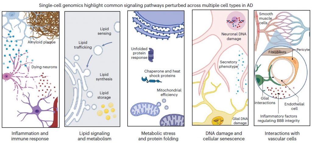
After decades of fundamental scientific and drug discovery research, Alzheimer’s disease has remained inscrutable and incurable, with a bare minimum of therapeutic progress. But in a new review article in Nature Neuroscience, MIT scientists write that by employing the new research capability of single-cell profiling, the field has rapidly achieved long-sought insights with strong potential for both explaining Alzheimer’s disease and doing something meaningful about it. By analyzing this new evidence, for instance, the authors show that the disease’s disruptions converge on five main areas of cellular function, or “pathways,” in each of five major brain cell types.

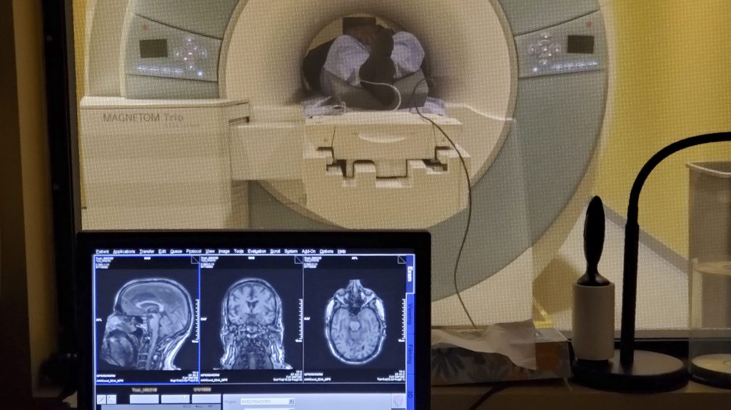
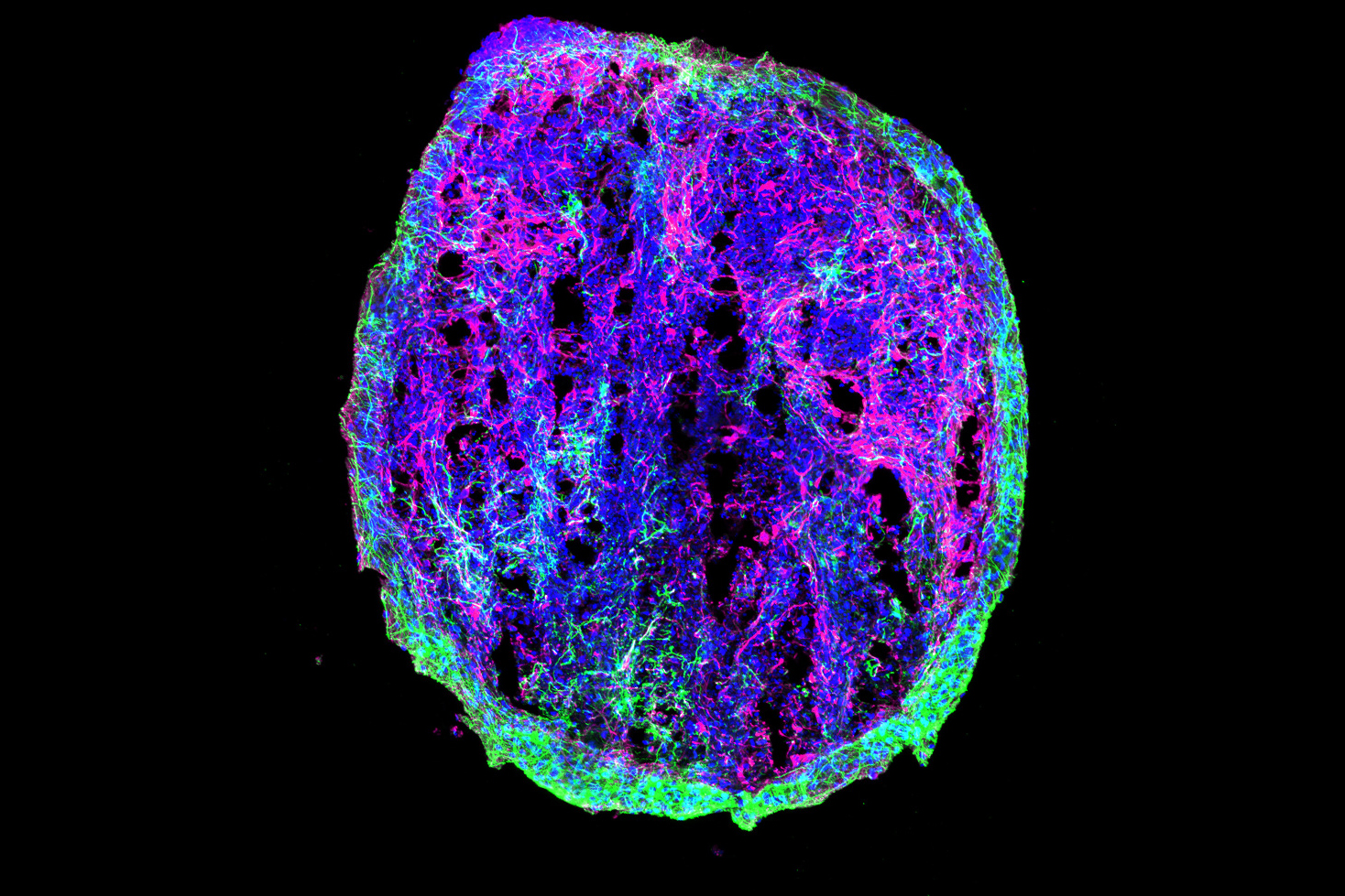
Single-cell profiling technologies produce comprehensive measurements of genetic activity in individual cells, such as levels of RNA which is transcribed from DNA, so that the cell’s functions and roles of in the biology of the brain, and the pathology of disease, can be assessed. Single-cell profiling technologies go beyond genome sequencing, which catalogs the DNA present in most every cell of a person, by revealing how each cell is uniquely making use of that common set of instructions. In studying Alzheimer’s disease, scientists have been using single-cell profiling to see how various brain cells, such as distinct types of neurons and microglia and astrocytes act differently in disease compared to how they behave in a healthy brain.

In the article, MIT Brain and Cognitive Sciences doctoral student Mitch Murdock and Picower Professor Li-Huei Tsai, Director of MIT’s Picower Institute for Learning and Memory and Aging Brain Initiative, write that while the findings of single-cell profiling studies confirm that the disease’s terrible effects are complex and far-reaching, there appear to also be five pathways that become perturbed in each of five major cell types. Investigating these pathways, they write, could produce valuable biomarkers of disease and yield meaningful targets for therapeutic intervention:

  • Inflammation and immune response
  • Lipid (fat molecule) signaling and metabolism
  • Metabolic stress and protein folding
  • DNA damage and cellular senescence (aging)
  • Interactions with brain vasculature (blood vessels)
Five panels offer cartoon schematics of a wide variety of cell and molecular interactions. The graphic is headed by this text: "single-cell genomics highlght common signaling pathways perturbed across multiple cell types in AD"
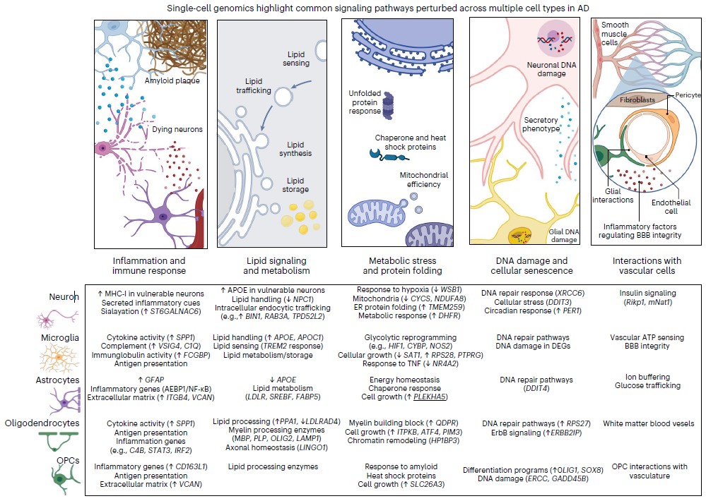
A figure from the paper depicts pathways perturbed in Alzheimer’s disease. Below examples are listed for each pathway by major brain cell type.

For each of these pathways in neurons, microglia, astrocytes, oligodendrocytes and oligodendrocyte precursor cells, Tsai and Murdock identify specific differences in gene regulation, found in single-cell studies, that significantly occur in brains of Alzheimer’s patients or mouse models compared to healthy control samples.

For example, Tsai and Murdock highlight more than a dozen genes all intimately involved in lipid processing whose expression is altered in various ways in various cells in the brain’s prefrontal cortex. For another example they show that all five cell types show impairments in DNA repair, albeit by changed expression of different genes in each.

“By identifying vulnerable cell types and the molecular programs that give rise to them, therapeutic interventions might reverse aberrant cellular trajectories,” Murdock and Tsai wrote in Nature Neuroscience. “While many transcriptional alterations are cell-type specific, these changes ultimately might converge on shared signaling pathways across cell types that might represent targets for new therapeutic strategies.”

To be sure, the authors note, there is still plenty of work to be done, both in refining and improving on single-cell techniques and also exploiting newer related opportunities. The paper notes a number of issues that must be carefully considered in producing valid single-cell profiling results, including where cells are sampled in the brain for sequencing, from whom, and in what condition. Moreover, it’s not always straightforward to show how changes in gene expression necessarily affect biology and it’s even harder to know whether any particular intervention, for instance to target altered inflammation pathways, will prove safe and effective as a therapy.

Future directions, meanwhile could include making greater use of “spatial transcriptomics,” which measures gene transcription in cells where they are situated within the brain, rather than removing them for analysis. Studies should be expanded to incorporate more human samples so that varying disease and demographic differences can be fully accounted for. Datasets should be shared and integrated, the authors write, and better comparisons between human and mouse samples are necessary to better understand how well, or not, they overlap.

“Single-cell profiling facilitates a nuanced portrait of the diverse cellular processes perturbed in the AD brain,” Tsai and Murdock conclude. “These varied molecular programs help explain the divergence between healthy aging and cognitive decline, and highlight cell-type-specific molecular programs involved in AD. Core signaling modules are disrupted across multiple cell types, and manipulating disrupted cellular states will pave the way for new therapeutic opportunities.”

The foreground shows a computer monitor with multiple views inside a brain. The background shows a person lying inside the tube of an MRI machine.

Small studies of 40Hz sensory stimulation confirm safety, suggest Alzheimer’s benefits

MIT researchers report early stage clinical study results of tests with non-invasive 40Hz light and sound treatment

A pair of early stage clinical studies testing the safety and efficacy of 40Hz sensory stimulation to treat Alzheimer’s disease has found that the potential therapy was well tolerated, produced no serious adverse effects and was associated with some significant neurological and behavioral benefits among a small cohort of participants.

“In these clinical studies we were pleased to see that volunteers did not experience any safety issues and used our experimental light and sound devices in their homes consistently,” said Li-Huei Tsai, Picower Professor in the The Picower Institute for Learning and Memory at MIT and senior author of the paper describing the studies in PLOS ONE Dec. 1. “While we are also encouraged to see some significant positive effects on the brain and behavior, we are interpreting them cautiously given our study’s small sample size and brief duration. These results are not sufficient evidence of efficacy, but we believe they clearly support proceeding with more extensive study of 40Hz sensory stimulation as a potential non-invasive therapeutic for Alzheimer’s disease.”

In three studies spanning 2016-2019, Tsai’s lab discovered that exposing mice to light flickering or sound clicking at the gamma-band brain rhythm frequency of 40Hz – or employing the light and sound together – produced widespread beneficial effects. Treated mice modeling Alzheimer’s disease pathology experienced improvements in learning and memory; reduced brain atrophy, neuron and synapse loss; and showed lower levels of the hallmark Alzheimer’s proteins amyloid beta and phosphorylated tau compared to untreated controls. The stimulation appears to produce these effects by increasing the power and synchrony of the 40Hz brain rhythm, which the lab has shown profoundly affects the activity of several types of brain cells, including the brain’s vasculature.Study designs

Based on those encouraging results, Diane Chan, a neurologist at Massachusetts General Hospital and a postdoctoral clinical fellow in Tsai’s lab, led the two new clinical studies at MIT. One set of tests, a “Phase 1” study, enrolled 43 volunteers of various ages including 16 people with early stage Alzheimer’s to confirm that exposure to 40Hz light and sound was safe and test whether it increased 40Hz rhythm and synchrony after a few minutes of exposure, as measured with EEG electrodes. The study also included two patients with epilepsy at the University of Iowa who consented to having measurements taken in deeper brain structures during exposure to 40Hz sensory stimulation while undergoing epilepsy-related surgery.

The second set of tests, a “Phase 2A” pilot study, enrolled 15 people with early stage Alzheimer’s disease in a single-blinded, randomized, controlled study to receive exposure to 40Hz light and sound (or non-40Hz “sham” stimulation for experimental controls) for an hour a day for at least three months. They underwent baseline and follow-up visits including EEG measurements during stimulation, MRI scans of brain volume, and cognitive testing. The stimulation device the volunteers used in their homes (a light panel synchronized with a speaker) was equipped with video cameras to monitor device usage. Participants also wore sleep-monitoring bracelets during their participation in the trial.

The Phase 2A trial launched just before the onset of the Covid-19 pandemic in 2020, causing some participants to become unable to undergo follow ups after three months. The study therefore only reports results through a four-month period.

Study results

In the Phase 1 study volunteers filled out a questionnaire on side effects, reporting a few minor but no major adverse effects. The most common was feeling “sleepy or drowsy.” Meanwhile, measurements taken with EEG scalp electrodes clustered at frontal and occipital sites showed significant increases in 40Hz rhythm power at each cortical site among cognitively normal younger and older participants as well as volunteers with mild Alzheimer’s. The readings also demonstrated significant increase in coherence at the 40Hz frequency between the two sites. Between the two volunteers with epilepsy, measurements showed significant increases in 40Hz power in deeper brain regions such as the gyrus rectus, amygdala, hippocampus and insula with no adverse events including seizures.

In the Phase 2A study, neither treated nor control volunteers reported serious adverse events. Both groups used their devices 90 percent of the time. The eight volunteers treated with 40Hz stimulation experienced several beneficial effects that reached statistical significance compared to the seven volunteers in the control condition. Control participants exhibited two signs of brain atrophy as expected with disease progression: reduced volume of the hippocampus and increased volume of open spaces, or ventricles. Treated patients did not experience significant changes in these measures. Treated patients also exhibited better connectivity across brain regions involved in the brain’s default mode and medial visual networks, which are related to cognition and visual processing respectively. Treated patients also exhibited more consistent sleep patterns than controls.

“Overall, these findings suggest that 40Hz GENUS has positive effects on AD-related pathology and symptoms and should be studied more extensively to evaluate its potential as a disease-modifying intervention for AD.”

Neither the treatment and control groups showed any differences after just three months on most cognitive tests, but the treatment group did perform significantly better on a face-name association test, a memory task with a strong visual component. The two groups, which were evenly matched by age, gender, APOE risk gene status, and cognitive scores, differed by years of education but that difference had no relationship to the results, the researchers wrote.

“After such a short time we didn’t expect to see significant effects on cognitive measures so it was encouraging to see that at least on face-name association the treatment group did perform significantly better,” Chan said.

In PLOS ONE the researchers concluded: “Overall, these findings suggest that 40Hz GENUS has positive effects on AD-related pathology and symptoms and should be studied more extensively to evaluate its potential as a disease-modifying intervention for AD.”

After the study ended all participants were permitted to continue using the devices set to provide the 40Hz stimulation.

The MIT team is now planning new clinical studies to test whether 40Hz sensory stimulation may be effective in preventing the onset of Alzheimer’s in high-risk volunteers and is launching preliminary studies to determine its therapeutic potential for Parkinson’s disease and Down syndrome. Cognito Therapeutics, an MIT spin-off company co-founded by Tsai and co-author Ed Boyden, Y. Eva Tan Professor of Neurotechnology at MIT, has launched Phase 3 trials of 40Hz sensory stimulation as an Alzheimer’s treatment using a different device.

Tsai, Boyden and co-author Emery N. Brown, Edward Hood Taplin Professor of Computational Neuroscience and Medical Engineering at MIT, are among the co-founders of MIT’s Aging Brain Initiative, which has advanced this collaboration and other neurodegeneration research at MIT.

In addition to Tsai, Chan, Boyden and Brown, the study’s other authors are Ho-Jun Suk, Brennan Jackson, Noah Milman, Danielle Stark, Elizabeth Klerman, Erin Kitchener, Vanesa S. Fernandez Avalos, Gabrielle de Weck, Arit Banerjee, Sara D. Beach, Joel Blanchard, Colton Stearns, Aaron D. Boes, Brandt Uitermarkt, Phillip Gander, Matthew Howard III, Eliezer J. Sternberg, Alfonso Nieto-Castanon, Sheeba Anteraper, Susan Whitfield-Gabrieli, and Bradford C. Dickerson.

Funding for the study came from sources including the Robert A. and Renee E. Belfer Family Foundation, Ludwig Family Foundation, JPB Foundation, Eleanor Schwartz Charitable Foundation, the Degroof-VM Foundation, Halis Family Foundation, and David B Emmes, Gary Hua and Li Chen, the Ko Han Family, Lester Gimpelson, Elizabeth K. and Russell L. Siegelman, Joseph P. DiSabato and Nancy E. Sakamoto, Alan and Susan Patricof, Jay L. and Carroll D Miller, Donald A. and Glenda G. Mattes, the Marc Haas Foundation, Alan Alda, and Dave Wargo.

A 3x2 array of squares shows a top row of three pinkish brain tissue samples with clear dark streaks. On the bottom row the pinkish squares of brain tissue don't show such a clear pattern

Alzheimer’s risk gene undermines insulation of brain’s “wiring”

In people carrying the APOE4 risk variant, a key brain cell type mismanages cholesterol needed to insulate neurons properly—another sign that APOE4 contributes to disease by disrupting lipids in the brain.

It’s well known that carrying one copy of the APOE4 gene variant increases one’s risk for Alzheimer’s disease threefold and two copies about tenfold, but the fundamental reasons why and what can be done to help patients remain largely unknown. A study published by an MIT-based team Nov. 16 in Nature provides some new answers as part of a broader line of research that has demonstrated APOE4’s consequences cell type by cell type in the brain.

The new study combines evidence from postmortem human brains, lab-based human brain cell cultures, and Alzheimer’s model mice to show that when people have one or two copies of APOE4, rather than the more common and risk-neutral APOE3 version, cells called oligodendrocytes mismanage cholesterol, failing to transport the fat molecule to wrap the long vine-like axon “wiring” that neurons project to make brain circuit connections. Deficiency of this fatty insulation, called myelin, may be a significant contributor to the pathology and symptoms of Alzheimer’s disease because without proper myelination, communications among neurons are degraded.

Recent studies by the research group, led by Picower Professor Li-Huei Tsai, director of The Picower Institute for Learning and Memory and the Aging Brain Initiative at MIT, have found distinct ways that APOE4 disrupts how fat molecules, or lipids, are handled by key brain cell types including neurons, astrocytes and microglia. In the new study as well as in those, the team has identified compounds that appear in the lab to correct these different problems, yielding potential pharmaceutical-based treatment strategies.

The new study extends that work not only by discovering how APOE4 disrupts myelination, but also by providing the first systematic analysis across major brain cell types using single nucleus RNA sequencing (snRNAseq) to compare how gene expression differs in people with APOE4 compared to APOE3.

“This paper shows very clearly from the snRNAseq of postmortem human brains in a genotype specific manner that APOE4 influences different brain cell types very distinctly,” said Tsai, a member of MIT’s Brain and Cognitive Sciences faculty. “We see convergence of lipid metabolism being disrupted, but when you really look into further detail at the kind of lipid pathways being disturbed in different brain cell types, they are all different.

“I feel that lipid dysregulation could be this very fundamental biology underlying a lot of the pathology we observe,” she said.

The paper’s lead authors are Joel Blanchard, an assistant professor at Mt. Sinai’s Icahn School of Medicine who began the work as a postdoc in Tsai’s MIT lab, Djuna Von Maydell and Leyla Akay, who are graduate students in Tsai’s lab, and Jose Davila Velderrain, a research group leader at Human Technopole and former postdoc in the lab of co-corresponding author Manolis Kellis, Computer Science Professor at MIT.

Many methods to examine myelination

Postmortem human brain samples came from the Religious Orders Study and the Rush Memory and Aging Project. The team’s snRNAseq results, a dataset that von Maydell has made freely available, encompasses more than 160,000 individual cells of 11 different types from the prefrontal cortex of 32 people—12 with two APOE3 copies, 12 with one copy of each APOE3 and APOE4, and eight with two APOE4 copies. The APOE3/3 and APOE3/4 samples were balanced by Alzheimer’s diagnosis, gender, and age. All APOE4/4 carriers had Alzheimer’s and 5 of 8 were female.

Some results reflected known Alzheimer’s pathology, but other patterns were novel. One in particular showed that APOE4-carrying oligodendrocytes exhibited greater expression of cholesterol synthesis genes and disruptions to cholesterol transport. The more APOE4 copies people had, the greater the effect. This was especially interesting given results from a prior analysis by Tsai’s and Kellis’s labs in 2019 that linked Alzheimer’s disease to reduced expression of myelination genes among oligodendrocytes.

Using a variety of techniques to look directly at the tissue, the team saw that in APOE4 brains, aberrant amounts of cholesterol accumulated within cell bodies, especially of oligodendrocytes, but was relatively lacking around neural axons.

To understand why, the team used patient-derived induced pluripotent stem cells to create lab cell cultures of oligodendrocytes engineered to differ only by whether they had APOE4 or APOE3. Again APOE4 cells showed major lipid disruptions. In particular, the afflicted oligodendrocytes hoarded extra cholesterol within their bodies, showed signs that the extra internal fats were stressing organelles called the endoplasmic reticulum that have a role in cholesterol transport, and indeed transported less cholesterol out to their membranes. Later when they were co-cultured with neurons, the APOE4 oligodendrocytes failed to myelinate the neurons as well as APO3 cells did, regardless of whether the neurons carried APOE4 or APOE3.

The team also observed that in post-mortem brains there was less myelination in APOE4 carriers than APOE3 carriers. For instance, the sheaths around axons running through the corpus callosum (the structure that connects brain hemispheres) were notably thinner in APOE4 brains. The same was true in mice engineered to harbor human APOE4 vs. those engineered to have APOE3.

A productive intervention

Eager to find a potential intervention, the team focused on drugs that affect cholesterol including statins (which suppress synthesis) and cyclodextrin, which aids cholesterol transport. The statins didn’t help, but applying cyclodextrin to APOE4 oligodendrocyte cultured in a dish reduced accumulation of cholesterol within the cells and improved myelination in co-cultures with neurons. Moreover, it also had these effects in APOE4 mice.

Finally, the team treated some APOE4 mice with cyclodextrin, left others untreated, and subjected them all to two different memory tests.  The cyclodextrin-treated mice performed both tests significantly better, suggesting an association between improved myelination and improved cognition.

Tsai said a clear picture is emerging in which intervening to correct specific lipid dysregulations by cell type could potentially help counteract APOE4’s contributions to Alzheimer’s pathology.

“It’s encouraging that we’ve seen a way to rescue oligodendrocyte function and myelination in lab and mouse models,” Tsai said. “But in addition to oligodendrocytes, we may also need to find clinically effective ways to take care of microglia, astrocytes, and vasculature to really combat the disease.”

In addition to the lead authors, Tsai and Kellis, the paper’s other authors are Hansruedi Mathys, Shawn Davidson, Audrey EffenbergerChi0yu Chen, Kristan Maner-Smith, Ihab Jahhar, Eric Orlund, Michael Bula, Emre Agbas, Ayesha Ng., Xueqiao Jiang, Martin Kahn, Cristina Blanco-Duque, Nicolas Lavoie, Liwang Liu, Ricardo Reyes, Yuan-Ta lin, Tak Ko, Lea R’Bibo, William Ralvenius, David Bennet and Hugh Cam.

The Robert A. and Renee E. Belfer Foundation, the JPB Foundation, the Carol and Gene Ludwig Family Foundation the Cure Alzheimer’s Fund and the National Insitutes of Health funded the study.

ON a black background the M-shaped coronal cross section of a mouse brain featurse patches of magenta and teal dots

With fractured genomes, Alzheimer’s neurons call for help

Study indicates that ailing neurons may instigate an inflammatory response from the brain’s microglia immune cells

A new study by researchers in The Picower Institute for Learning and Memory at MIT provides evidence from both mouse models and postmortem human tissue of a direct link between two problems that emerge in Alzheimer’s disease: a buildup of double-stranded breaks (DSBs) in the DNA of neurons and the inflammatory behavior of microglia, the brain’s immune cells.

A key new finding is that neurons actively trigger an inflammatory response to their genomic damage. Neurons have not been known to signal the brain’s immune system in Alzheimer’s disease, said study lead author Gwyneth Welch, a former MIT Brain and Cognitive Sciences graduate student in the lab of senior author Li-Huei Tsai.

“This is a novel concept in neuroscience: the idea that neurons can be activating inflammatory activity in response to DNA damage,” Welch said. “The general idea was that neurons have a more passive relationship with microglia regarding age-associated neuroinflammation.”

Instead, what Welch, Tsai and co-authors report in Science Advances is that neurons coping with mounting DSBs go through stages of first trying to fix their fractured DNA and then, when it apparently fails, sending out via molecular signals to microglia, which responded by taking on a more inflammatory state. In experiments where the scientists interrupted the immune signaling, they prevented microglia from entering that state and degrading neural circuit connections, or synapses.

Members of Tsai’s lab have been studying DSBs in the context of Alzheimer’s for more than a decade. Tsai said the new findings add to the emerging understanding of the role they play in Alzheimer’s.

“We have a longstanding interest in understanding DNA breaks in neurons,” said Tsai, Picower Professor of Neuroscience and a founder of MIT’s Aging Brain Initiative. “We previously showed that DNA double stranded breaks are necessary for the induction of activity-regulated gene expression in neurons but we also observed profound DNA damage in neurons in the early stages of neurodegeneration.

“We now know that DNA-damaged neurons exhibit senescent phenotypes and play an active role in eliciting an immune response from microglia and perhaps astrocytes,” Tsai said. “This is mediated by the activation of the NFkappaB transcription factor. Moreover, we identified two cytokines secreted by damaged neurons to recruit microglia and elicit microglial response.  Importantly, we show that inhibition of NFkappaB rescued synaptic loss in neurodegeneration, further elucidating of impact of neuroimmune response on synaptic integrity and cognitive function.”

In her thesis research Welch used the lab’s “CK-p25” mouse model of Alzheimer’s, in which disease pathology can be induced, She observed a timeline in which neurons with DSBs appeared within a week, peaked in number after two weeks and then tapered off, becoming notably reduced by six weeks. Meanwhile, those neurons also lost their ability to express a standard marker of neuronal identity. Welch realized there seemed to be stages to the process of coping with DSBs. First neurons have few DSBs and strong identity (baseline), then high DSBs with no loss of identity (stage 1), then high DSBs and a loss of neuronal identity (stage 2).

Transcripts tell the tale

To understand what cells were doing differently in each stage, Welch and the team used multiple “transcriptomics” technologies, which tracks differences in gene expression. Her analyses revealed that neuronal identity genes were most strongly expressed at baseline, DNA repair genes were strong during stage 1, and immune signaling genes were particularly prominent during stage 2.

Among the immune signaling genes were ones governed by the master transcription regulator NFkappaB. These included the cytokines Ccl2 and Cxcl10.

To see if these changes were due specifically to DSBs, Welch treated neurons in the absence of any induced pathology with a chemical called etoposide that causes DSBs. Similar gene expression patterns evident in the induced mice were recapitulated in the ones treated with etoposide. And when Welch also looked at gene expression in the brains of people with DSBs and with Alzheimer’s she also found many significant overlaps.

“We found that stage 1 and stage 2 gene signatures were active in human DSB-bearing neurons,” she and her co-authors wrote. “This neuronal immune signature was further amplified in the context of AD pathology, suggesting that it may serve a functional role in disease-associated neuroinflammation.”

A big role for microglia

Having established that DSB-afflicted neurons employ NFkappaB to send out immune signals such as Ccl2 and Cxcl10, Welch and the team then asked what the effect was. Given that the lab in 2017 had characterized a late-stage inflammatory response on the part of microglia in Alzheimer’s, they hypothesized that neurons might be responsible.

For this analysis Welch used spatial transcriptomics. She divided up both uninduced and induced mouse brains into many areas, and rated each area based on the strength of their DSB signal. Then she analyzed gene transcription in each area and found that locations with high DSBs also had many more microglia in an inflammatory state than locations with low DSBs. They were also able to directly image this relationship, yielding the observation that inflammatory microglia (evidenced by abnormally large cell bodies) were co-located with high-DSB neurons.

To further test the hypothesis, they disrupted NFkappaB regulated transcription in neurons by interfering with a key molecular cog in that machinery called p65. That step resulted in reduced proliferation of microglia and reduced microglia cell body size. It also induced beneficial changes in microglia gene expression, making them more consistent with their normal “homeostatic” state.

In other experiments they found that etoposide-treated neurons expressed Cxcl10 and Ccl2 but that disrupting NFkappaB reduced that expression. They also saw that depleting the two molecules from the brains also prevented microglia from becoming reactive.

And looking back at the neurons, they saw that while knocking down NFkappaB activity didn’t prevent them from dying, it did preserve circuit connections, or synapses, on the neurons that remained alive. That’s important because those circuit connections underlie brain function and microglia are known to prune those.

Because NFkappaB is known to help prevent cell death (which may be why knocking it down didn’t prevent neurons from dying), Welch said knocking it down is not likely to be a therapeutic strategy.

“Its more a proof of principle that that if you turn off a major switch for inflammation, that will change how microglia and neurons interact,” she said. “If your goal is to target inflammatory pathways, focusing on specific signaling molecules might be the more precise way to intervene.”

In addition to Welch and Tsai, the paper’s other authors are Carles Boix, Eloi Schmauch, Jose Davila-Velderrain, Matheus Victor, Vishnu Dileep, P. Lorenzo Bozzelli, Qiao Su, Jemmie D. Cheng, Audrey Lee, Noelle S. Leary, Andreas R. Pfenning, and Manolis Kellis.

The National Institutes of Health, CureAlz, the Glenn Foundation, and the JPB Foundation provided funding for the research.

A blue-hued cross-secction of a mouse;s brain highlights the visual cortex in red on the upper left and green on the upperright.

When Alzheimer’s degrades cells that cross hemispheres, visual memory suffers

New research reveals cells that span brain hemispheres to coordinate activity in visual processing centers and shows that Alzheimer’s degrades their structure and therefore their function

A new MIT study finds that Alzheimer’s disease disrupts at least one form of visual memory by degrading a newly identified circuit that connects the vision processing centers of each brain hemisphere.

The results of the study, published in Neuron by a research team based at The Picower Institute for Learning and Memory, come from experiments in mice, but provide a physiological and mechanistic basis for prior observations in human patients: the degree of diminished brain rhythm synchrony between counterpart regions in each hemisphere correlates with the clinical severity of dementia.

“We demonstrate that there is a functional circuit that can explain this phenomenon,” said lead author Chinnakkaruppan Adaikkan, a former Picower Institute postdoc who is now an assistant professor in the Centre for Brain Research at the Indian Institute of Science (IISc) in Bangalore. “In a way we uncovered a fundamental biology that was not known before.”

Specifically, Adaikkan’s work identified neurons that connect the primary visual cortex (V1) of each hemisphere and showed that when the cells are disrupted, either by genetic alterations that model Alzheimer’s disease or by direct laboratory perturbations, brain rhythm synchrony becomes reduced and mice become significantly less able to notice when a new pattern appeared on a wall in their enclosures. Such recognition of novelty, which requires visual memory of what was there the prior day, is an ability commonly disrupted in Alzheimer’s.

“This study demonstrates the propagation of gamma rhythm synchrony across the brain hemispheres via the cross hemispheric connectivity,” said study senior author Li-Huei Tsai, Picower Professor and director of The Picower Institute and MIT’s Aging Brain Initiative. “It also demonstrates that the disruption of this circuit in AD mouse models is associated with specific behavioral deficits.”

Cross-hemispheric cells

In the study, Adaikkan, Tsai, Thomas McHugh and co-authors discovered and traced V1 neurons that extended their axons all the way through the corpus callosum, which connects the brain’s hemispheres, to cells in the V1 on the brain’s other side. There, they found, the cross-hemispheric (CH) neurons forged connections, or synapses, with target cells, providing them with “excitatory” stimulation to drive their activity. Adaikkan also found that CH neurons were much more likely to be activated by a novelty discrimination task than V1 neurons in general or neurons in other regions heavily involved in memory such as the hippocampus or the prefrontal cortex.

Curious about how this might differ in Alzheimer’s disease, the team looked at the activity of the cells in two different Alzheimer’s mouse models. The found that CH cell activity was significantly lessened amid the disease. Unsurprisingly, Alzheimer’s mice fared much poorer in novelty discrimination tasks.

The team examined the CH cells closely and found that they gather incoming input from a variety of other cells within their V1 and other regions in their hemisphere that process visual information. When they compared the incoming connections of healthy CH neurons to those in CH cells afflicted with Alzheimer’s, they found that cells in the disease condition had significantly less infrastructure for hosting incoming connections (measured in terms of synapse-hosting spines protruding from the vine-like dendrites that sprawl out of the cell body).

Given the observations correlating reduced brain rhythm synchrony and memory performance in Alzheimer’s, the team wondered if that occurred in the mice, too. To find out, they custom-designed electrodes to measure rhythmic activity simultaneously in all cortical layers of each hemisphere’s V1. They observed that cross-hemispheric synchrony increased notably between the V1s when mice engaged in novelty discrimination but that the synchrony, both at high “gamma” and lower “theta” frequency rhythms, was significantly lower in the Alzheimer’s mice than it was in healthy mice.

Adaikkan’s evidence at that point was strong, but still only suggestive, that CH neurons provided the means by which the V1 regions on each side of the brain could coordinate to enable novelty discrimination, and that this ability became undermined by Alzheimer’s degradation of the CH cells’ connectivity. To more directly determine whether the CH circuit played such a causal, consequential role, the team directly intervened to disrupt them, testing what effect targeted perturbations had.

They found that chemically inhibiting CH cells disrupted rhythm synchrony between V1s, mirroring measures made in Alzheimer’s model mice. Moreover, disrupting CH activity undermined novelty discrimination ability. To further test whether it was the cells’ cross-hemispheric nature that mattered specifically, they engineered CH cells to be controllable with flashes of light (a technology called “optogenetics”). When they shined the light on the connections, they forged in the other hemisphere to inhibit those, they found that doing so again compromised visual discrimination ability.

All together, the study results show that CH cells in V1 connect with neurons in the counterpart area of the opposite hemisphere to synchronize neural activity needed for properly recognizing novelty, but that Alzheimer’s disease damages their ability to do that job.

Adaikkan said he is curious to now look at other potential cross-hemispheric connections and how they may be affected in Alzheimer’s disease, too. He said he also wants to study what happens to synchrony at other rhythm frequencies.

In addition to Adaikkan and Tsai, the study’s other authors are Jun Wang, Karim Abdelall, Steven Middleton, Lorenzo Bozzelli, and Ian Wickersham.

The JPB Foundation, The National Institutes of Health, and the Robert A. and Renee E. Belfer Foundation provided funding for the study.

A swirling blue ball of long thin neurons is overlaid with long electrodes coming in from the bottom and the left.

How microglia contribute to Alzheimer’s disease

A breakdown of lipid metabolism in these brain cells promotes inflammation and interferes with neuron activity, a new study finds

One of the hallmarks of Alzheimer’s disease is a reduction in the firing of some neurons in the brain, which contributes to the cognitive decline that patients experience. A new study from MIT shows how a type of cells called microglia contribute to this slowdown of neuron activity.

The study found that microglia that express the APOE4 gene, one of the strongest genetic risk factors for Alzheimer’s disease, cannot metabolize lipids normally. This leads to a buildup of excess lipids that interferes with nearby neurons’ ability to communicate with each other.

“APOE4 is a major genetic risk factor, and many people carry it, so the hope is that by studying APOE4, that will also provide a bigger picture of the fundamental pathophysiology of Alzheimer’s disease and what fundamental cell processes that have to go wrong to result in Alzheimer’s disease,” says Li-Huei Tsai, director of MIT’s Picower Institute for Learning and Memory and the senior author of the study.

On a black background we see a nearly round mass of tangly blue neurons with flecks of green throughout
For their study researchers cultured spheroids of neurons. Here neurons are stained purple among more general cell nuclei (blue) and astrocyte cells (green).

The findings suggest that if researchers could find a way to restore normal lipid metabolism in microglia, that might help to treat some of the symptoms of the disease.

MIT postdoc Matheus Victor is the lead author of the paper, which appears today in Cell Stem Cell.

Lipid overload

About 14 percent of the population has the APOE4 variant, making it the most common genetic variant that has been linked to late-onset, nonfamilial Alzheimer’s disease. People who carry one copy of APOE4 have a threefold higher risk of developing Alzheimer’s, and people with two copies have a tenfold higher risk.

“If you look at this another way, if you look at the entire Alzheimer’s disease population, about 50 percent of them are APOE4 carriers. So, it’s a very significant risk, but we still don’t know why this APOE4 allele presents such a risk,” Tsai says.

The APOE gene also comes in two other forms, known as APOE2, which is considered protective against Alzheimer’s, and the most common form, APOE3, which is considered neutral. APOE3 and APOE4 differ by just one amino acid.

For several years, Tsai’s lab has been studying the effects of APOE4 on a variety of cell types in the brain. To do this, the researchers use induced pluripotent stem cells, derived from human donors, and engineer them to express a specific version of the APOE gene. These cells can then be stimulated to differentiate into brain cells, including neurons, microglia, and astrocytes.

In a 2018 study, they showed that APOE4 causes neurons to produce large quantities of amyloid beta peptide 42, an Alzheimer’s-linked molecule that causes the neurons to become hyperactive. That study found that APOE4 also affects the functions of microglia and astrocytes, leading to cholesterol accumulation, inflammation, and failure to clear amyloid beta peptides.

A 2021 follow-up showed that APOE4 astrocytes have dramatic impairments in their ability to process a variety of lipids, which leads to a buildup of molecules such as triglycerides, as well as cholesterol. In that paper, the researchers also showed that treating engineered yeast cells expressing APOE4 with choline, a dietary supplement that is a building block for phospholipids, could reverse many of the detrimental effects of APOE4.

In their new study, the researchers wanted to investigate how APOE4 affects interactions between microglia and neurons. Recent research has shown that microglia play an important role in modulating neuronal activity, including their ability to communicate within neural ensembles. Microglia also scavenge the brain looking for signs of damage or pathogens, and clear out debris.

The researchers found that APOE4 disrupts microglia’s ability to metabolize lipids and prevents them from removing lipids from their environment. This leads to a buildup of fatty molecules, especially cholesterol, in the environment. These fatty molecules bind to a specific type of potassium channel embedded in neuron cell membranes, which suppresses neuron firing.

“We know that in late stages of Alzheimer’s disease, there is reduced neuron excitability, so we may be mimicking that with this model,” Victor says.

The buildup of lipids in microglia can also lead to inflammation, the researchers found, and this type of inflammation is believed to contribute to the progression of Alzheimer’s disease.

Restoring function

The researchers also showed that they could reverse the effects of lipid overload by treating APOE4 microglia with a drug called Triacsin C, which interferes with the formation of lipid droplets. When APOE4 microglia were exposed to this drug, the researchers found that normal communication between neighboring neurons and microglia was restored.

“We can rescue the suppression of neuronal activity by APOE4 microglia, presumably through lipid homeostasis being restored, where now fatty acids are not accumulating extracellularly,” Victor says.

Triacsin C can be toxic to cells, so it would likely not be suitable to use as a drug to treat Alzheimer’s, but the researchers hope that other approaches to restore lipid homeostasis could help combat the disease. In Tsai’s 2021 APOE4 study, she showed that choline also helps to restore normal microglia activity.

“Lipid homeostasis is actually critical for a number of cell types across the Alzheimer’s disease brain, so it’s not singularly a microglia problem,” Victor says. “The question is, how do you restore lipid homeostasis across multiple cell types? It’s not an easy task, but we’re tackling that through choline, for example, which might be a really interesting angle.”

The researchers are now further studying how microglia transition from a healthy state to a “lipid-burdened,” inflammatory state, in hopes of discovering ways to block that transition. In previous studies in mice, they have shown that exposure to LED light flickering at a specific frequency can help to rejuvenate microglia, stimulating the cells to resume their normal functions.

The research was funded by the National Institutes of Health, the Howard Hughes Medical Institute Hanna H. Gray Postdoctoral Fellowship, The Robert A. and Renee E. Belfer Family Foundation, Carol and Eugene Ludwig Family Foundation, the Cure Alzheimer’s Fund, The JPB Foundation, Joseph P. DiSabato and Nancy E. Sakamoto, Donald A. and Glenda G. Mattes, Lester A. Gimpelson, the Halis Family Foundation, the Dolby family, David Emmes, and Alan and Susan Patricof.

–From MIT News
Three bright yellow-green celll cultures on black background

In Down syndrome cells, genome-wide disruptions mimic a senescence-like state

Extra chromosome alters chromosomal conformation and DNA accessibility across the whole genome in neural progenitor cells, disrupting gene transcription and cell functions much like in cellular aging

In Down syndrome, the third copy of chromosome 21 causes a reorganization of the 3D configuration of the entire genome in a key cell type of the developing brain, a new study shows. The resulting disruption of gene transcription and cell function are so similar to those seen in cellular aging, or senescence, that the scientists leading the study found they could use anti-senescence drugs to correct them in cell cultures.

The study published in Cell Stem Cell therefore establishes senescence as a potentially targetable mechanism for future treatment of Down syndrome, said Hiruy Meharena, a new assistant professor at the University of California San Diego who led the work as a Senior Alana Fellow in the Alana Down Syndrome Center at MIT.

“There is a cell-type specific genome-wide disruption that is independent of the gene dosage response,” Meharena said. “It’s a very similar phenomenon to what’s observed in senescence. This suggests that excessive senescence in the developing brain induced by the third copy of chromosome 21 could be a key reason for the neurodevelopmental abnormalities seen in Down syndrome.”

The study’s finding that neural progenitor cells (NPCs), which develop into major cells in the brain including neurons, have a senescent character is remarkable and novel, said senior author Li-Huei Tsai, but it is substantiated by the team’s extensive work to elucidate the underlying mechanism of the effects of abnormal chromosome number, or aneupoloidy, within the nucleus of the cells.

“This study illustrates the importance of asking fundamental questions about the underlying mechanisms of neurological disorders,” said Tsai, Picower Professor of Neuroscience, director of the Alana Center, and of The Picower Institute for Learning and Memory at MIT. “We didn’t begin this work expecting to see senescence as a translationally relevant feature of Down syndrome, but the data emerged from asking how the presence of an extra chromosome affects the architecture of all of a cell’s chromosomes during development.”

Genomewide changes

Meharena and co-authors spent years measuring distinctions between human cell cultures that differed only by whether they had a third copy of chromosome 21. Stem cells derived from volunteers were cultured to turn into NPCs. In both the stem cells and the NPCs, the team examined 3D chromosome architecture, several metrics of DNA structure and interaction, gene accessibility and transcription, and gene expression. They also looked at the consequences of the gene expression differences on important functions of these developmental cells, such as how well they proliferated and migrated in 3D brain tissue cultures. Stem cells were not particularly different, but NPCs were substantially affected by the third copy of chromosome 21.

Li-Huei Tsai stands in a lab and points to an image of a cell on a computer screen. Hiruy Meharena sits at the desk with his hand on the mouse and looks on.
Li-Huei Tsai and Hiruy Meharena consult about images produced during the research in this 2019 photo

Overall, the picture that emerged in NPCs was that the presence of a third copy causes all the other chromosomes to squish inward, not unlike when people in a crowded elevator must narrow their stance when one more person squeezes in. The main effects of this “chromosomal introversion,” meticulously quantified in the study, are more genetic interactions within each chromosome and less interactions among them. These changes and differences in DNA conformation within the cell nucleus lead to changes in how genes are transcribed and therefore expressed, causing important differences in cell function that affect brain development.

Treated as senescence

For the first couple of years as these data emerged, Meharena said, the full significance of the genomic changes were not apparent, but then he read a paper showing very similar genomic rearrangement and transcriptional alterations in senescent cells.

After validating that the Down syndrome cells indeed bore such a similar signature of transcriptional differences, the team decided to test whether anti-senescence drugs could undo the effects. They tested a combination of two: dasatinib and quercetin. The medications improved not only gene accessibility and transcription, but also the migration and proliferation of cells.

That said, the drugs have very significant side effects—dasatinib is only given to cancer patients when other treatments have not done enough—so they are not appropriate for attempting to intervene in brain development amid Down syndrome, Meharena said. Instead an outcome of the study could be to inspire a search for medications that could have anti-senolytic effects with a safer profile.

A schematic shows the process of how a trisomy 21 cell experiences genomiwide chromosomal reconfiguration leading to a senescence-like response
A new study’s findings indicate that amid trisomy 21, neural progenitor cells experience a genome-wide chromosomal reorganization leading to a senescence-like response including altered chromatin states and gene transcription.

Senescence is a stress response of cells. At the same time, years of research by former MIT biology professor Angelika Amon, who co-directed the Alana Center with Tsai, has shown that aneuploidy is a source of considerable stress for cells. A question raised by the new findings, therefore, is whether the senescence-like character of Down syndrome NPCs is indeed the result of an aneuploidy induced stress and if so, exactly what that stress is.

Another implication of the findings is how excessive senescence among brain cells might affect people with Down syndrome later in life. The risk of Alzheimer’s disease is much higher at a substantially earlier age in the Down syndrome population than among people in general. In large part this is believed to be because a key Alzheimer’s risk gene, APP, is on chromosome 21, but the newly identified inclination for senescence may also accelerate Alzheimer’s development.

In addition to Meharena and Tsai, the paper’s other authors are Asaf Marco, Vishnu Dileep, Elana Lockshin, Grace Akatsu, James Mullahoo, Ashley Watson, Tak Ko, Lindsey Guerin, Fatema Abdurrob, Shruti Rengarajan, Malvina Papanastasiou and Jacob Jaffe.

The Alana Foundation, the LuMind Foundation, Burroughs Wellcome Fund, UNCF-Merck  and the National Institutes of Health funded the research.

Boston Globe op-ed: How science, technology, and industry can work together to cure Alzheimer’s

The Alzheimer’s research community must acknowledge the gaps in the current approach to curing the disease and make significant changes.

In The Boston Globe Nov. 29, Li-Huei Tsai penned an op-ed as part of Globe’s “The Longevity Hub” collaboration with MIT Professor Joe Coughlin.

“Alzheimer’s disease, the sixth leading cause of death in the United States, has defied our best efforts to find a cure or even a treatment that can substantially slow its devastating degradation of the brain. The now decades-long sequence of high-profile setbacks in Alzheimer’s drug discovery and development underscores the unique challenge this disease presents.

For the sake not only of tens of millions of patients and families worldwide, but also the sustainability of the US health care system and economy, the Alzheimer’s research community must acknowledge the gaps in the current approach to curing the disease and make significant changes in how science, technology, and industry work together to meet this challenge…”

Read the full Op-Ed in the Globe here..


Study links gene to cognitive resilience in the elderly

The findings may help explain why some people who lead enriching lives are less prone to Alzheimer’s and age-related dementia

Many people develop Alzheimer’s or other forms of dementia as they get older. However, others remain sharp well into old age, even if their brains show underlying signs of neurodegeneration.

Among these cognitively resilient people, researchers have identified education level and amount of time spent on intellectually stimulating activities as factors that help prevent dementia. A new study by MIT researchers shows that this kind of enrichment appears to activate a gene family called MEF2, which controls a genetic program in the brain that promotes resistance to cognitive decline.

The researchers observed this link between MEF2 and cognitive resilience in both humans and mice. The findings suggest that enhancing the activity of MEF2 or its targets might protect against age-related dementia

“It’s increasingly understood that there are resilience factors that can protect the function of the brain,” says Li-Huei Tsai, director of MIT’s Picower Institute for Learning and Memory. “Understanding this resilience mechanism could be helpful when we think about therapeutic interventions or prevention of cognitive decline and neurodegeneration-associated dementia.”


Above: When researchers knocked down a Mef2 gene in the frontal cortex of mice (blank areas), it prevented the mice from attaining a cognitive benefit of being raised in an enriched environment.


Tsai, a founding director of MIT’s Aging Brain Initiative, is the senior author of the study, which appears today in Science Translational Medicine. The lead authors are recent MIT PhD recipient Scarlett Barker and MIT postdoctoral fellow and Boston Children’s Hospital physician Ravikiran (Ravi) Raju.

Scarlett Barker stands at a podium with audience members sitting in the foreground.
Co-lead author Scarlett Barker delivered remarks about the research at Neuroscience 2019 while it was in progress.

Protective effects

A large body of research suggests that environmental stimulation offers some protection against the effects of neurodegeneration. Studies have linked education level, type of job, number of languages spoken, and amount of time spent on activities such as reading and doing crossword puzzles to higher degrees of cognitive resilience.

The MIT team set out to try to figure how these environmental factors affect the brain at the neuronal level. They looked at human datasets and mouse models in parallel, and both tracks converged on MEF2 as a critical player.

MEF2 is a transcription factor that was originally identified as a factor important for cardiac muscle development, but later was discovered to play a role in neuron function and neurodevelopment. In two human datasets comprising slightly more than 1,000 people all together, the MIT team found that cognitive resilience was highly correlated with expression of MEF2 and many of the genes that it regulates.

Many of those genes encode ion channels, which control a neuron’s excitability, or how easily it fires an electrical impulse. The researchers also found, from a single-cell RNA-sequencing study of human brain cells, that MEF2 appears to be most active in a subpopulation of excitatory neurons in the prefrontal cortex of resilient individuals.

To study cognitive resilience in mice, the researchers compared mice who were raised in cages with no toys, and mice placed in a more stimulating environment with a running wheel and toys that were swapped out every few days. As they found in the human study, MEF2 was more active in the brains of the mice exposed to the enriched environment. These mice also performed better in learning and memory tasks.

When the researchers knocked out the gene for MEF2 in the frontal cortex, this blocked the mice’s ability to benefit from being raised in the enriched environment, and their neurons became abnormally excitable.

“This was particularly exciting as it suggested that MEF2 plays a role in determining overall cognitive potential in response to variables in the environment,” Raju says.

Ravi Raju stands at a podium. A blue and white Picower Institute logo is behind him as a backdrop.
Co-lead author Ravikiran (Ravi) Raju presents some of his early research during a 2018 talk. Image by Adrienne Mathiowetz.

The researchers then explored whether MEF2 could reverse some of the symptoms of cognitive impairment in a mouse model that expresses a version of the tau protein that can form tangles in the brain and is linked with dementia. If these mice were engineered to overexpress MEF2 at a young age, they did not show the usual cognitive impairments produced by the tau protein later in life. In these mice, neurons overexpressing MEF2 were less excitable.

“A lot of human studies and mouse model studies of neurodegeneration have shown that the neurons become hyperexcitable in early stages of disease progression,” Raju says. “When we overexpressed MEF2 in a mouse model of neurodegeneration, we saw that it was able to prevent this hyperexcitability, which might explain why they performed cognitively better than control mice.”

Enhancing resilience

The findings suggest that enhancing MEF2 activity could help to protect against dementia; however, because MEF2 also affects other types of cells and cellular processes, more study is needed to make sure that activating it wouldn’t have adverse side effects, the researchers say.

The MIT team now hopes to further investigate how MEF2 becomes activated by exposure to an enriching environment. They also plan to examine some of the effects of the other genes that MEF2 controls, beyond the ion channels they explored in this study. Such studies could help to reveal additional targets for drug treatments.

“You could potentially imagine a more targeted therapy by identifying a subset or a class of effectors that is critically important for inducing resilience and neuroprotection,” Raju says.

The research was funded by the Glenn Center for Biology of Aging Research, the National Institute of Aging, the Cure Alzheimer’s Fund, and the Eunice Kennedy Shriver National Institute of Child Health and Human Development.

-From MIT News

A colorful cartoon of a DNA double helix with a gap through the middle

Memory making involves extensive DNA breaking

To quickly express genes needed for learning and memory, brain cells snap both strands of DNA in many more places and cell types than previously realized, a new study shows

The urgency to remember a dangerous experience requires the brain to make a series of potentially dangerous moves: Neurons and other brain cells snap open their DNA in numerous locations—more than previously realized, according to a new study—to provide quick access to genetic instructions for the mechanisms of memory storage.

The extent of these DNA double-strand breaks (DSBs) in multiple key brain regions is surprising and concerning, said study senior author Li-Huei Tsai, Picower Professor of Neuroscience at MIT and director of The Picower Institute for Learning and Memory, because while the breaks are routinely repaired, that process may become more flawed and fragile with age. Tsai’s lab has shown that lingering DSBs are associated with neurodegeneration and cognitive decline and that repair mechanisms can falter.

“We wanted to understand exactly how widespread and extensive this natural activity is in the brain upon memory formation because that can give us insight into how genomic instability could undermine brain health down the road,” said Tsai, who is also a professor in the Department of Brain and Cognitive Sciences and a leader of MIT’s Aging Brain Initiative. “Clearly memory formation is an urgent priority for healthy brain function but these new results showing that several types of brain cells break their DNA in so many places to quickly express genes is still striking.”

Tracking breaks

In 2015, Tsai’s lab provided the first demonstration that neuronal activity caused DSBs and that they induced rapid gene expression. But those findings, mostly made in lab preparations of neurons, did not capture the full extent of the activity in the context of memory formation in a behaving animal and did not investigate what happened in cells other than neurons.

IIn the new study published July 1 in PLOS ONE, lead author and former graduate student Ryan Stott and co-author and former research technician Oleg Kritsky sought to investigate the full landscape of DSB activity in learning and memory. To do so, they gave mice little electrical zaps to the feet when they entered a box, to condition a fear memory of that context. They then used several methods to assess DSBs and gene expression in the brains of the mice over the next half hour, particularly among a variety of cell types in the prefrontal cortex and hippocampus, two regions essential for the formation and storage of conditioned fear memories. They also made measurements in the brains of mice who did not experience the foot shock to establish a baseline of activity for comparison.

The creation of a fear memory doubled the number of DSBs among neurons in the hippocampus and the prefrontal cortex, affecting more than 300 genes in each region. Among 206 affected genes common to both regions, the researchers then looked at what those genes do. Many were associated with the function of the connections neurons make with each other, called synapses. This makes sense because learning arises when neurons change their connections (a phenomenon called “synaptic plasticity”) and memories are formed when groups of neurons connect together into ensembles called engrams.

“Many genes essential for neuronal function and memory formation, and significantly more of them than expected based on previous observations in cultured neurons…are potentially hotspots of DSB formation,” the authors wrote in the study.

In another analysis, the researchers confirmed through measurements of RNA that the increase in DSBs indeed correlated closely with increased transcription and expression of affected genes, including ones affecting synapse function, as quickly as 10-30 minutes after the foot shock exposure.

“Overall, we find transcriptional changes are more strongly associated with [DSBs] in the brain than anticipated,” they wrote. “Previously we observed 20 gene-associated [DSB] loci following stimulation of cultured neurons, while in the hippocampus and prefrontal cortex we see more than 100-150 gene associated [DSB] loci that are transcriptionally induced.”

Snapping with stress

In the analysis of gene expression, the neuroscientists looked at not only neurons but also non-neuronal brain cells, or glia, and found that they also showed changes in expression of hundreds of genes after fear conditioning. Glia called astrocytes are known to be involved in fear learning, for instance, and they showed significant DSB and gene expression changes after fear conditioning.

Among the most important functions of genes associated with fear conditioning-related DSBs in glia was the response to hormones. The researchers therefore looked to see which hormones might be particularly involved and discovered that it was glutocortocoids, which are secreted in response to stress. Sure enough, the study data showed that in glia, many of the DSBs that occurred following fear conditioning occurred at genomic sites related to glutocortocoid receptors. Further tests revealed that directly stimulating those hormone receptors could trigger the same DSBs that fear conditioning did and that blocking the receptors could prevent transcription of key genes after fear conditioning.

Tsai said the finding that glia are so deeply involved in establishing memories from fear conditioning is an important surprise of the new study.

“The ability of glia to mount a robust transcriptional response to glutocorticoids suggest that glia may have a much larger role to play in the response to stress and its impact on the brain during learning than previously appreciated,” she and her co-authors wrote.

Damage and danger?

More research will have to be done to prove that the DSBs required for forming and storing fear memories are a threat to later brain health, but the new study only adds to evidence that it may be the case, the authors said.

“Overall we have identified sites of DSBs at genes important for neuronal and glial functions, suggesting that impaired DNA repair of these recurrent DNA breaks which are generated as part of brain activity could result in genomic instability that contribute to aging and disease in the brain,” they wrote.

The National Institutes of Health, The Glenn Foundation for Medical Research and the JPB Foundation provided funding for the research.