Category: Uncategorized

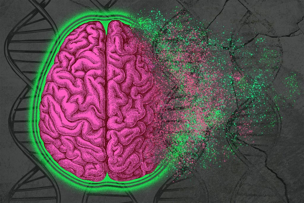
A pink cartoon brain is superposed over a background of DNA double-helixes. Toward the right side of the image, the brain is crumbling to dust.

Decoding the complexity of Alzheimer’s disease

By analyzing epigenomic and gene expression changes that occur in Alzheimer’s disease, researchers identify cellular pathways that could become new drug targets

Alzheimer’s disease affects more than 6 million people in the United States, and there are very few FDA-approved treatments that can slow the progression of the disease.

In hopes of discovering new targets for potential Alzheimer’s treatments, MIT researchers have performed the broadest analysis yet of the genomic, epigenomic, and transcriptomic changes that occur in every cell type in the brains of Alzheimer’s patients.

Using more than 2 million cells from more than 400 postmortem brain samples, the researchers analyzed how gene expression is disrupted as Alzheimer’s progresses. They also tracked changes in cells’ epigenomic modifications, which help to determine which genes are turned on or off in a particular cell. Together, these approaches offer the most detailed picture yet of the genetic and molecular underpinnings of Alzheimer’s.

The researchers report their findings in a set of four papers appearing today in Cell. The studies were led by Li-Huei Tsai, director of MIT’s Picower Institute for Learning and Memory, and Manolis Kellis, a professor of computer science in MIT’s Computer Science and Artificial Intelligence Laboratory (CSAIL) and a member of the Broad Institute of MIT and Harvard.

“What we set out to do was blend together our computational and our biological expertise and take an unbiased look at Alzheimer’s at an unprecedented scale across hundreds of individuals — something that has just never been undertaken before,” Kellis says.

The findings suggest that an interplay of genetic and epigenetic changes feed on each other to drive the pathological manifestations of the disease.

“It’s a multifactorial process,” said Tsai, who leads MIT’s Aging Brain Initiative. “These papers together use different approaches that point to a converging picture of Alzheimer’s disease where the affected neurons have defects in their 3D genome, and that is causal to a lot of the disease phenotypes we see.”

A complex interplay

Many efforts to develop drugs for Alzheimer’s disease have focused on the amyloid plaques that develop in patients’ brains. In their new set of studies, the MIT team sought to uncover other possible approaches by analyzing the molecular drivers of the disease, the cell types that are the most vulnerable, and the underlying biological pathways that drive neurodegeneration.

To that end, the researchers performed transcriptomic and epigenomic analyses on 427 brain samples from the Religious Orders Study/Memory and Aging Project (ROSMAP), a longitudinal study that has tracked memory, motor, and other age-related changes in older people since 1994. These samples included 146 people with no cognitive impairment, 102 with mild cognitive impairment, and 144 diagnosed with Alzheimer’s-linked dementia.

In the first Cell paper, focused on gene expression changes, the researchers used single-cell RNA-sequencing to analyze the gene expression patterns of 54 types of brain cells from these samples, and identified cellular functions that were most affected in Alzheimer’s patients. Among the most prominent, they found impairments in the expression of genes involved in mitochondrial function, synaptic signaling, and protein complexes needed to maintain the structural integrity of the genome.

This gene expression study, which was led by former MIT postdoc Hansruedi Mathys, graduate student Zhuyu (Verna) Peng, and former graduate student Carles Boix, also found that genetic pathways related to lipid metabolism were highly disrupted. In work published in Nature last year, the Tsai and Kellis labs showed that the strongest genetic risk for Alzheimer’s, called APOE4, interferes with normal lipid metabolism, which can then lead to defects in many other cell processes.

In the study led by Mathys, the researchers also compared gene expression patterns in people who showed cognitive impairments and those who did not, including some who remained sharp despite having some degree of amyloid buildup in the brain, a phenomenon known as cognitive resilience. That analysis revealed that cognitively resilient people had larger populations of two subsets of inhibitory neurons in the prefrontal cortex. In people with Alzheimer’s-linked dementia, those cells appear to be more vulnerable to neurodegeneration and cell death.

“This revelation suggests that specific inhibitory neuron populations might hold the key to maintaining cognitive function even in the presence of Alzheimer’s pathology,” Mathys says. “Our study pinpoints these specific inhibitory neuron subtypes as a crucial target for future research and has the potential to facilitate the development of therapeutic interventions aimed at preserving cognitive abilities in aging populations.”

Epigenomics

In the second Cell paper, led by former MIT postdoc Xushen Xiong, graduate student Benjamin James, and former graduate student Carles Boix PhD ’22, the researchers examined some of the epigenomic changes that occurred in 92 people, including 48 healthy individuals and 44 with early or late-stage Alzheimer’s. Epigenomic changes are alterations in the chemical modifications or packaging of DNA that affect the usage of a particular gene within a given cell.

To measure those changes, the researchers used a technique called ATAC-Seq, which measures the accessibility of sites across the genome at single-cell resolution. By combining this data with single-cell RNA-sequencing data, the researchers were able to link information about how much a gene is expressed with data on how accessible that gene is. They could also start to group genes into regulatory circuits that control specific cell functions such as synaptic communication — the primary way that neurons transmit messages throughout the brain.

Using this approach, the researchers were able to track changes in gene expression and epigenomic accessibility that occur in genes that have previously been linked with Alzheimer’s. They also identified the types of cells that were most likely to express these disease-linked genes, and found that many of them occur most often in microglia, the immune cells responsible for clearing debris from the brain.

This study also revealed that every type of cell in the brain undergoes a phenomenon known as epigenomic erosion as Alzheimer’s disease progresses, meaning that the cells’ normal pattern of accessible genomic sites is lost, which contributes to loss of cell identity.

The role of microglia

In a third Cell paper, led by MIT graduate student Na Sun and research scientist Matheus Victor, the researchers focused primarily on microglia, which make up 5 to 10 percent of the cells in the brain. In addition to clearing debris from the brain, these immune cells also respond to injury or infection and help neurons communicate with each other.

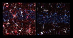
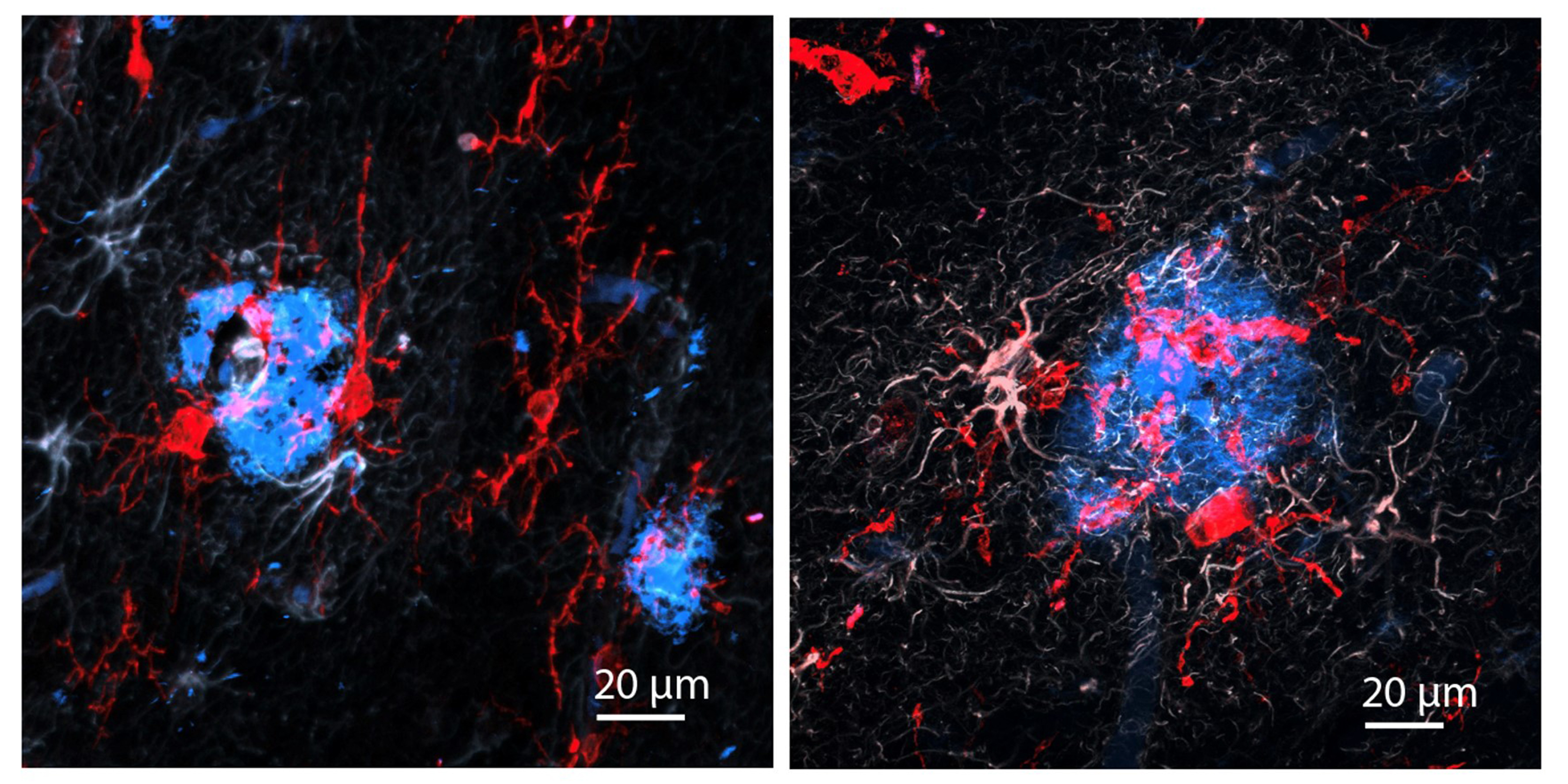
In two panels red microglia surround a bluish blob. In the left panel the microglia are more slender whereas they are more bloated on the right. The blue blub is more compact on the left and more diffuse on the right.
Researchers tracked changes in microglia early (left) and late (right) in Alzheimer’s disease. Microglia (red) surround an amyloid plaque (blue). The microglia are noticeably more activated, with larger cell bodies, on the right. The amyloid plaque, meanwhile, is more diffused. Images by Mat Victor.

This study builds on a 2015 paper from Tsai and Kellis in which they found that many of the genome-wide association study (GWAS) variants associated with Alzheimer’s disease are predominantly active in immune cells like microglia, much more than in neurons or other types of brain cells.

In the new study, the researchers used RNA sequencing to classify microglia into 12 different states, based on hundreds of genes that are expressed at different levels during each state. They also showed that as Alzheimer’s disease progresses, more microglia enter inflammatory states. The Tsai lab has also previously shown that as more inflammation occurs in the brain, the blood-brain barrier begins to degrade and neurons begin to have difficulty communicating with each other.

At the same time, fewer microglia in the Alzheimer’s brain exist in a state that promotes homeostasis and helps the brain function normally. The researchers identified transcription factors that turn on the genes that keep microglia in that homeostatic state, and the Tsai lab is now exploring ways to activate those factors, in hopes of treating Alzheimer’s disease by programming inflammation-inducing microglia to switch back to a homeostatic state.

DNA damage

In the fourth Cell study, led by MIT research scientist Vishnu Dileep and Boix, the researchers examined how DNA damage contributes to the development of Alzheimer’s disease. Previous work from Tsai’s lab has shown that DNA damage can appear in neurons long before Alzheimer’s symptoms appear. This damage is partly a consequence of the fact that during memory formation, neurons create many double-stranded DNA breaks. These breaks are promptly repaired, but the repair process can become faulty as neurons age.

This fourth study found that as more DNA damage accumulates in neurons, it becomes more difficult for them to repair the damage, leading to genome rearrangements and 3D folding defects.

“When you have a lot of DNA damage in neurons, the cells, in their attempt to put the genome back together, make mistakes that cause rearrangements,” Dileep says. “The analogy that I like to use is if you have one crack in an image, you can easily put it back together, but if you shatter an image and try to piece it back together, you’re going to make mistakes.”

These repair mistakes also lead to a phenomenon known as gene fusion, which occurs when rearrangements take place between genes, leading to dysregulation of genes. Alongside defects in genome folding, these changes appear to predominantly impact genes related to synaptic activity, likely contributing to the cognitive decline seen in Alzheimer’s disease.

The findings raise the possibility of seeking ways to enhance neurons’ DNA repair capabilities as a way to slow down the progression of Alzheimer’s disease, the researchers say.

In addition, Kellis’ lab now hopes to use artificial intelligence algorithms such as protein language models, graph neural networks, and large language models to discover drugs that might target some of the key genes that the researchers identified in these studies.

The researchers also hope that other scientists will make use of their genomic and epigenomic data. “We want the world to use this data,” Kellis says. “We’ve created online repositories where people can interact with the data, can access it, visualize it, and conduct analyses on the fly.”

The research was funded, in part, by the National Institutes of Health and the Cure Alzheimer’s Foundation CIRCUITS consortium.

Research papers:
–Story by MIT News. Lead image by Christine Daniloff
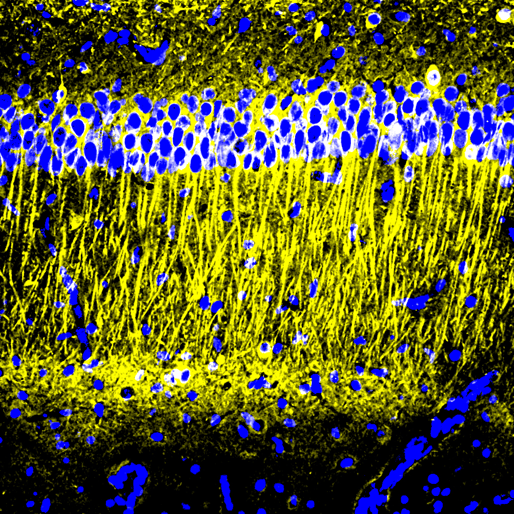
A square panel shows tall yellow streaks. At the very bottom is a black field with a few blue dots. Almost across the top there is a strip of many blue dots.

Molecule reduces inflammation in Alzheimer’s models

A potential new Alzheimer’s drug represses the harmful inflammatory response of the brain’s immune cells, reducing disease pathology, preserving neurons and improving cognition in preclinical tests

Though drug developers have achieved some progress in treating Alzheimer’s disease with medicines that reduce amyloid-beta protein, other problems of the disease including inflammation, continue unchecked. In a new study, scientists at The Picower Institute for Learning and Memory at MIT describe a candidate drug that in human cell cultures and Alzheimer’s mouse models reduced inflammation and improved memory.

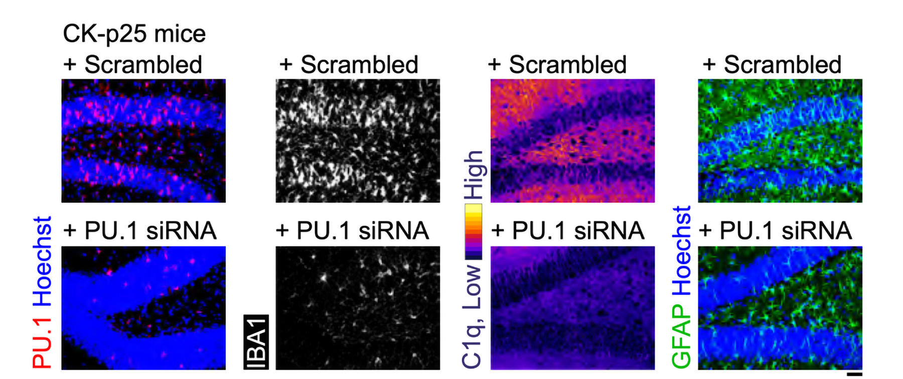
The target of the new “A11” molecule is a genetic transcription factor called PU.1. Prior research has shown that amid Alzheimer’s disease, PU.1 becomes an overzealous director of inflammatory gene expression in the brain’s microglia immune cells. A11 suppresses this problematic PU.1 activity, the new research shows, by recruiting other proteins that repress the inflammatory genes PU.1 works to express. But because A11 concentrates mostly in the brain and does not reduce PU.1 levels, it does not appear to disrupt PU.1’s other job, which is to ensure the production of a wide variety of blood cells.

“Inflammation is a major component of Alzheimer’s disease pathology that has been especially hard to treat,” said study senior author Li-Huei Tsai, Picower Professor of Neuroscience at MIT and director of The Picower Institute and MIT’s Aging Brain Initiative. “This preclinical study demonstrates that A11 reduces inflammation in human microglia-like cells as well as in multiple mouse models of Alzheimer’s disease and significantly improves cognition in the mice. We believe A11 therefore merits further development and testing.”

Tsai and Elizabeta Gjoneska of the National Institutes of Health are co-corresponding authors of the study published in the Journal of Experimental Medicine.

As a postdoc, Gjoneska co-led a 2015 study that implicated PU.1 as a regulator of errant microglia inflammation in a mouse model of Alzheimer’s disease. That research was a collaboration between Tsai’s lab and that of MIT Computer Science Professor Manolis Kellis, co-led by former postdoc Andreas Pfenning, now a faculty member at Carnegie Mellon University. Ever since then, Tsai has been seeking a safe way to restore PU.1 activity to healthier levels.

The work described in the new paper, led by Picower Institute research scientist William Ralvenius, starts with experiments to further validate that PU.1 would be a therapeutically meaningful target. To do that the scientists compared gene expression in immune cells of postmortem brain samples from Alzheimer’s patients and mouse models and matching non-Alzheimer’s controls. The comparisons showed that Alzheimer’s effects major changes in microglial gene expression and that an increase in PU.1 binding to inflammatory gene targets was a significant component of that change. Moreover, they showed that reducing PU.1 activity in a mouse model of Alzheimer’s reduced inflammation and neurodegeneration, the death of neurons.

Screening success

Genetically knocking down PU.1 in the body is not a viable therapeutic strategy given its importance in normal healthy function. The team therefore screened more than 58,000 small molecules from libraries of FDA-approved drugs and novel chemicals to see if any could safely and significantly reduce key inflammation and Alzheimer’s related genes regulated by by PU.1 in cell cultures. After several rounds of increasingly stringent screening, they narrowed the field down to six chemicals. A11 was by far the most potent among them.

They tested the effects of A11 doses on the function of human microglia-like cells cultured from patient stem cells. When they exposed the microglia-like cells to immune molecules that typically trigger inflammation, cells dosed with A11 exhibited reduced expression and secretion of inflammatory cytokines and less of the cell body shape changes associated with microglia inflammatory responses. The cells also showed less accumulation of lipid molecules, another sign of inflammatory activation. Looking at gene expression patterns, the scientists observed that A11-treated cells exposed to inflammatory triggers behaved much like unperturbed microglia, suggesting that A11 helps prevent microglia from overreacting to inflammatory cues.

Two more lab tests aimed at understanding how A11 exerts its effects revealed that it doesn’t change PU.1 levels. Instead it counteracts PU.1 activity by recruiting several proteins including MECP2, HDAC1, SIN3A and DMNT3A, known to repress expression of its targets. Essentially amid Alzheimer’s disease, A11 tamps down what PU.1 amps up.

“A11 represents a first-in-class molecule that converts PU.1 from a transcriptional activator to a transcriptional repressor, resulting in a controlled state of microglial inflammation,” the authors wrote.

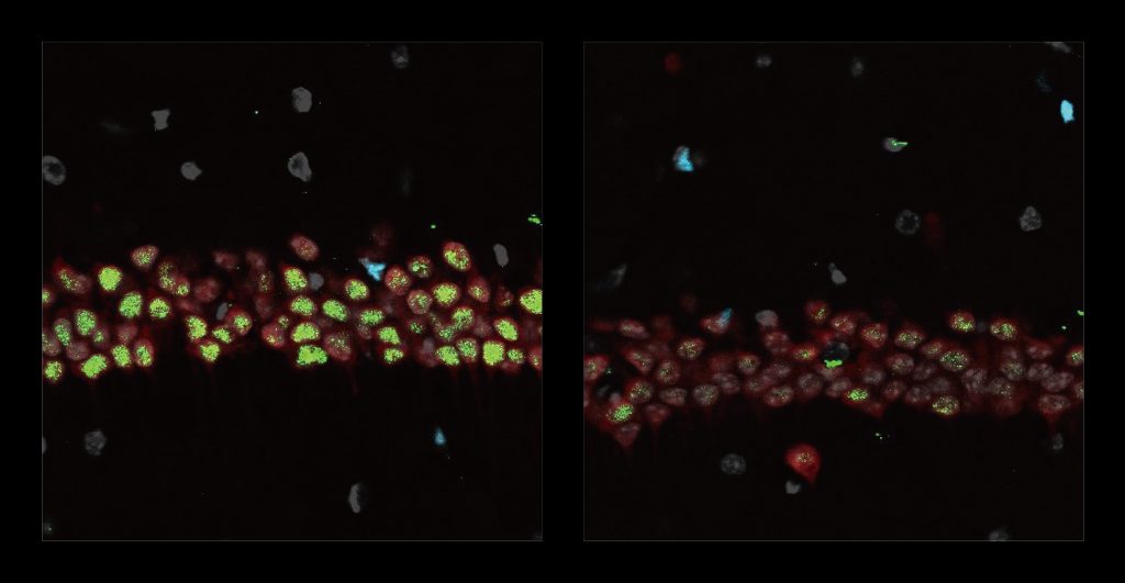
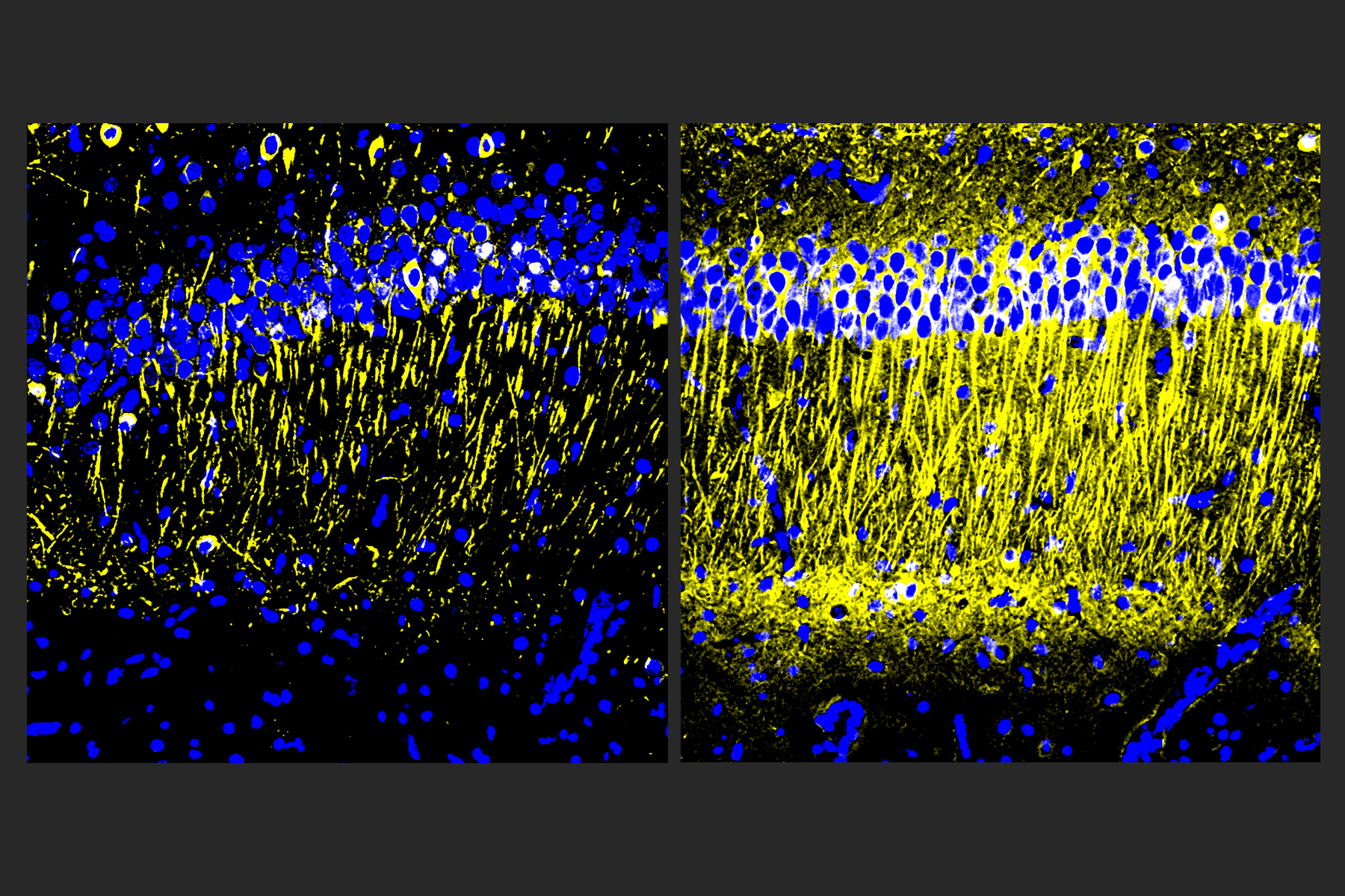
Two square panels side by side show a stripe of many blue dots near the top. The panel on the left has a few yellow stripes extending down from the blue dots. The panel on the right has many more of the yellow vertical lines.
The brains of Alzheimer’s model mice treated with A11 (right) showed more tubulin (yellow), a marker of neuronal health, than untreated controls (left).


Mice in mazes

Having established that A11 reduced inflammatory activity in microglia and determined how that happens, the team focused on whether it worked as a medicine in mouse models of Alzheimer’s disease.

Pharmacological tests indicated that A11 is readily cleared from tissues and is capable of reaching brain cells.  Moreover, in healthy mice the chemical successfully crossed the blood-brain barrier and remained in brain cells much longer than anywhere else.

Finally the team tested the effects of the drugs on Alzheimer’s disease pathology and symptoms in three mouse strains that each model different aspects of Alzheimer’s disease: CK-p25 mice (severe neurodegeneration), Tau P301S transgenic mice (tauopathy), and 5XFAD mice (amyloid pathology).

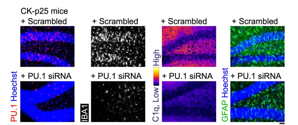
Male and female CK-p25 mice dosed with A11 showed less inflammatory response among microglia and astrocyte cells and lost fewer neurons than untreated controls. TauP301S Tg mice responded similarly, also exhibiting a significant reduction of phosphorylated tau protein in the hippocampus region of the brain, which is an essential area for memory. In 5XFAD mice, amyloid was significantly reduced.

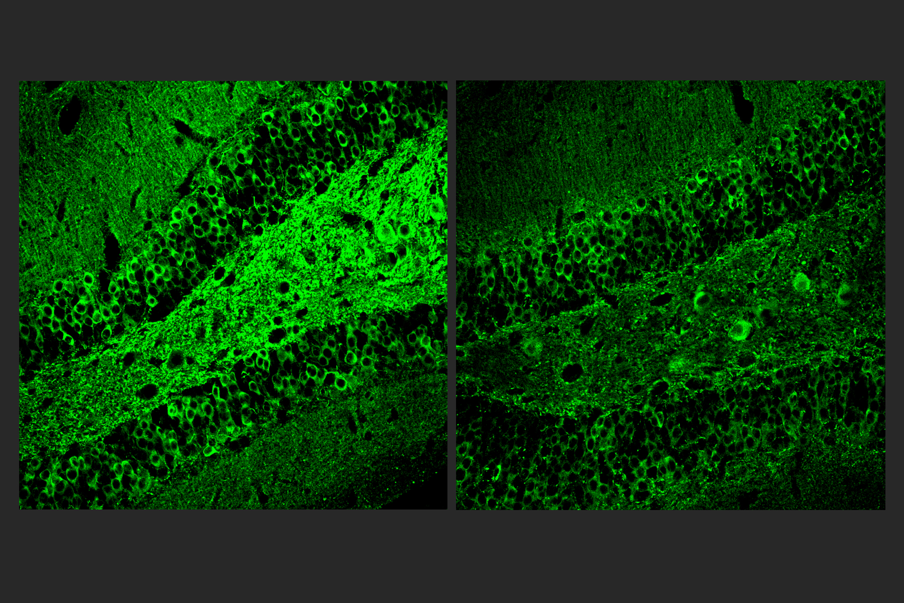
Two square panels side by side show brain tissue with green staining. The panel on the left shows much more green than the panel on the right.
A11 treated Alzheimer’s model mice (right) showed much less tau (green staining) than untreated controls (left).


The team subjected the Tau P301S Tg and CK-p25 mice to mazes designed to test their short-term working memory and longer-term learning. In both models and on both tests, A11-treated mice performed significantly better than untreated controls. For example, in the “Morris Water Maze,” where mice have to learn the location of a submerged platform that allows them to rest, treated CK-p25 mice learned much faster than untreated ones.

Much more testing needs to be done before A11 could become an approved medicine, Tsai said, but she noted that it could complement the new treatments that target amyloid.

“Given that A11 acts via a distinct mechanism from existing AD therapeutics, A11 could be used alone or in combination with approved therapeutics to provide improved treatment options for neurodegenerative diseases,” the authors concluded.

In addition to Tsai, Gjoneska and Ralvenius, the paper’s other authors are Alison E. Mungenast, Hannah Woolf, Margaret M. Huston, Tyler Z. Gillingham, Stephen K. Godin, Jay Penney, Hugh P. Cam, Fan Gao, Celia G. Fernandez, Barbara Czako, Yaima Lightfoot, William J. Ray, Adrian Beckmann, Alison M. Goate, Edoardo Marcora, Carmen Romero-Molina, Pinar Ayata, and Anne Schaefer.

The Robert A. and Renee E. Belfer Family Foundation and the National Institutes of Health funded the research. Additional support came from The JPB Foundation and The Picower Institute for Learning and Memory, The Halis Family Foundation, Lester A. Gimpelson and Jay L. and Caroll Miller.