Category: Neuroscience

Chinna Adaikkan stands before a research poster and talks to two onlookers.

Scientists eager to explain brain rhythm boost’s broad impact in Alzheimer’s models

The sweeping extent to which increasing 40Hz “gamma” rhythm power in the brain can affect the pathology and symptoms of Alzheimer’s disease in mouse models has been surprising, even to the MIT neuroscientists who’ve pioneered the idea. So surprising, in fact, they can’t yet explain why it happens.

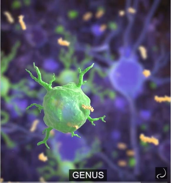
In three papers, including two this year in Cell and Neuron, they’ve demonstrated that exposing mice to light flickering or sound buzzing at 40Hz, a method dubbed “GENUS” for Gamma ENtrainment Using Sensory stimuli, strengthens the rhythm across the brain and changes the gene expression and activity of multiple brain cell types. Pathological amyloid and tau protein buildups decline, neurons and their circuit connections are protected from degeneration and learning and memory endure significantly better than in disease model mice who do not receive GENUS.

In a new review article in Trends in Neurosciences two researchers leading those efforts lay out the few knowns and many unknowns that must be understood to determine how the widespread effects take place. It’s a challenge they relish because the answers could both break new scientific ground and help them improve how GENUS could become a therapeutic or preventative approach for people.

“While we know it affects pathology in mice, we want to understand how because that will help us understand and refine potential treatment,” said lead author Chinnakkaruppan Adaikkan, a postdoc in the lab of senior author Li-Huei Tsai, Picower Professor of Neuroscience and director of The Picower Institute for Learning and Memory.

Adaikkan has been interested in understanding how neural activity produces brain rhythms since his doctoral research. At MIT, he is channeling that passion into understanding how sensory stimulation can entrain oscillations.

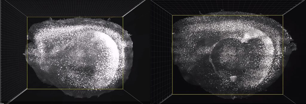
“That’s what drives me to come to the lab every day to study these mechanisms,” Adaikkan said. “When we got the data from the first mouse where we recorded from the visual cortex, the hippocampus and the prefrontal cortex we were surprised to see that visual stimulation entrains in these brain regions. That was very exciting but we have a very long way to go to understand how this happens.”

The new paper raises that question and many others for the field. What cells underlie the brain’s response to GENUS? How do gamma rhythms engage non-neuronal cells such as astrocytes and microglia? How does it propagate beyond the brain regions responsible for perception? How extensively can enhancing gamma affect cognition? Does long-term stimulation affect brain circuit connections and how they change?

Cell roles

Studies of how groups of neurons engage in coherent oscillations of electrical activity have yielded two models to explain gamma rhythms. Both involve an interplay between excitatory and inhibitory neurons but differ on which type leads the interaction, Adaikkan and Tsai wrote. In his work, Adaikkan is attempting to dissect the roles of specific neuron types in GENUS and how closely those patterns mirror other sources of gamma, such as that invoked by cognitive tasks.

GENUS affects more than neurons. Tsai’s lab has found that microglia change their gene expression, their physical form, their protein-consuming behavior and their inflammatory response depending on the Alzheimer’s model involved. Work from another group showed that blocking vesicle release in astrocytes can hinder gamma power in mice and Tsai’s group found that auditory GENUS recruits an increase reactive astrocytes, which are more inclined to consume pathological proteins.

The new paper offers three hypotheses about how such “glial” cells are involved: They might contribute directly to gamma entrainment by regulating the flow of ions that carry electrical charge; even if they don’t contribute to rhythms, their ionic sensitivity may still make them responsive to gamma changes; they might instead be affected by changes in levels of neurotransmitters as a result of gamma.

Moreover, different glia may also become involved because of their proximity to electrical couplings between neurons called synapses, or because of how their activity is otherwise governed by neural activity.

The broader brain

That GENUS extends to the hippocampus, which is key for memory, and the prefrontal cortex, which is key for cognition, is likely a factor in how it preserves brain function. But again there are competing models for how increased gamma could facilitate multi-regional communication. In one, the authors write, coherence at the same frequency optimizes communication, while in the other model, one region’s gamma activity directly drives activity in regions downstream. New experiments that directly manipulate inter-regional circuits, they argue, could help resolve which model better explains gamma entrainment’s effects.

Finally, the effects of GENUS on brain function and behavior also aren’t fully explained. The Tsai lab’s has shown significant effects on spatial memory and some effects on other forms of memory, depending on the stimulation method. Other studies have shown that stimulating brain rhythms by other means, such as via genetic or optogenetic manipulations in mice, or via transcranial stimulation in humans, can also improve functions such as working memory. Adaikkan is interested in closing a gap between those studies and the Tsai lab’s work: Most studies measure cognitive performance during stimulation, while the Tsai lab has done so after the conclusion of repeated stimulation. He said he’d like to also test how mice perform while GENUS is actively underway.

“Our lab is excited to tackle these many hypotheses and to see how the field tackles many more,” Tsai said. “GENUS has created many intriguing new questions for neuroscience.”

The JPB Foundation, The Robert A. and Renee E. Belfer Foundation, and the Jeffrey and Nancy Halis Family Foundation have supported the work.

Li-Huei Tsai is shown in profile as she stands smiling at a podium

Tsai elected fellow of National Academy of Inventors

The National Academy of Inventors has selected MIT neuroscientist Li-Huei Tsai, Picower Professor of Neuroscience and director of The Picower Institute for Learning and Memory, as a member of its 2019 class of new fellows.

NAI fellows “have demonstrated a highly prolific spirit of innovation in creating or facilitating outstanding inventions that have made a tangible impact on the quality of life, economic development and welfare of society,” the organization stated in its announcement.

Tsai’s research focuses on neurodegenerative conditions such as Alzheimer’s disease. Her work has generated a dozen patents, many of which have been licensed by biomedical companies including two startups, Cognito Therapeutics and Souvien Bio Ltd., that have spun out from her and collaborator’s labs. Her team’s innovations include inhibiting an enzyme that affects the chromatin structure of DNA to rescue gene expression and restore learning and memory, and using light and sound stimulation to enhance the power and synchrony of 40Hz gamma rhythms in the brain to reduce Alzheimer’s pathology, prevent neuron death and preserve learning and memory. Each of these promising sets of findings in mice are now being tested in human trials.

“The goal of my lab is to improve our understanding of neurodegenerative disease mechanisms and to develop new therapies that could prevent the suffering of millions of people and their loved ones,” said Tsai who also directs MIT’s Aging Brain Initiative. “A crucial part of that effort is translating promising fundamental findings to the clinic and I’m honored that the NAI has recognized our work toward that goal.”

Tsai joins 22 colleagues from MIT as an NAI fellow, including Materials Science and Engineering Department Head Christopher Schuh, who was also elected this year. Among previously elected fellows are Tsai collaborators Emery N. Brown, a fellow Picower Institute faculty member and Edward Hood Taplin Professor of Computational Neuroscience and Health Sciences & Technology, and Ed Boyden, Y. Eva Tan Professor of Neurotechnology.

A microscope image showing white dots of amyloid built up in a cluster within a mouse brain

Study pinpoints Alzheimer’s plaque emergence early and deep in the brain

Long before symptoms like memory loss even emerge, the underlying pathology of Alzheimer’s disease, such as an accumulation of amyloid protein plaques, is well underway in the brain. A longtime goal of the field has been to understand where it starts so that future interventions could begin there.

A new study by MIT neuroscientists at The Picower Institute for Learning and Memory could help those efforts by pinpointing the regions with the earliest emergence of amyloid in the brain of a prominent mouse model of the disease. Notably, the study also shows that the degree of amyloid accumulation in one of those same regions of the human brain correlates strongly with the progression of the disease.

“Alzheimer’s is a neurodegenerative disease so in the end you can see a lot of neuron loss,” said Wen-Chin “Brian” Huang, co-lead author of the study and a postdoc in the lab of co-senior author Li-Huei Tsai, Picower Professor of Neuroscience and director of the Picower Institute. “At that point it would be hard to cure the symptoms. It’s really critical to understand what circuits and regions show neuronal dysfunction early in the disease. This will in turn facilitate the development of effective therapeutics.”

In addition to Huang, the study’s co-lead authors are Rebecca Canter, a former member of the Tsai lab, and Heejin Choi, a former member of the lab of co-senior author Kwanghun Chung, associate professor of chemical engineering and a member of the Picower Institute and the Institute for Medical Engineering and Science.

Tracking plaques

Many research groups have made progress in recent years by tracing amyloid’s path in the brain using technologies such as positron emission tomography and by looking at brains post-mortem, but the new study in Communications Biology adds substantial new evidence from the 5XFAD mouse model because it presents an unbiased look at the entire brain as early as one month of age. The study reveals that amyloid begins its terrible march in deep brain regions such as the mammillary body, the lateral septum and the subiculum before making its way along specific brain circuits that ultimately lead it to the hippocampus, a key region for memory, and the cortex, a key region for cognition.

The team used SWITCH, a technology developed by Chung, to label amyloid plaques and to clarify the whole brains of 5XFAD mice so that they could be imaged in fine detail at different ages. The team was consistently able to see that plaques first emerged in the deep brain structures and then tracked along circuits such as the Papez memory circuit to spread throughout the brain by 6-12 months (a mouse’s lifespan is up to three years).

The findings help to cement an understanding that has been harder to obtain from human brains, Huang said, because post-mortem dissection cannot easily account for how the disease developed over time and PET scans don’t offer the kind of resolution the new study provides from the mice.

Key validations

Importantly, the team directly validated a key prediction of their mouse findings in human tissue: If the mammillary body is indeed a very early place that amyloid plaques emerge, then the density of those plaques should increase in proportion with how far advanced the disease is. Sure enough, when the team used SWITCH to examine the mammillary bodies of post-mortem human brains at different stages of the disease, they saw exactly that relationship: The later the stage, the more densely plaque-packed the mammillary body was.

“This suggests that human brain alterations in Alzheimer’s disease look similar to what we observe in mouse,” the authors wrote. “Thus we propose that amyloid-beta deposits start in susceptible subcortical structures and spread to increasingly complex memory and cognitive networks with age.”

The team also performed experiments to determine whether the accumulation of plaques they observed were of real disease-related consequence for neurons in affected regions. One of the hallmarks of Alzheimer’s disease is a vicious cycle in which amyloid makes neurons too easily excited and overexcitement causes neurons to produce more amyloid. The team measured the excitability of neurons in the mammillary body of 5XFAD mice and found they were more excitable than otherwise similar mice that did not harbor the 5XFAD set of genetic alterations.

In a preview of a potential future therapeutic strategy, when the researchers used a genetic approach to silence the neurons in the mammillary body of some 5XFAD mice but left neurons in others unaffected, the mice with silenced neurons produced less amyloid.

While the study findings help explain much about how amyloid spreads in the brain over space and time, they also raise new questions, Huang said. How might the mammillary body affect memory and what types of cells are most affected there?

“This study sets a stage for further investigation of how dysfunction in these brain regions and circuits contributes to the symptoms of Alzheimer’s disease,” he said.

In addition to Huang, Canter, Choi, Tsai and Chung, the paper’s other authors are Jun Wang, Lauren Ashley Watson, Christine Yao, Fatema Abdurrob, Stephanie Bousleiman, Jennie Young, David Bennett and Ivana Dellalle.

The National Institutes of Health, the JPB Foundation, Norman B. Leventhal and Barbara Weedon fellowships, The Burroughs Wellcome Fund, the Searle Scholars Program, a Packard Award, a NARSAD Young Investigator Award and the NCSOFT Cultural Foundation funded the research.

An animation shows a microglia cell gobbling an amyloid protein

New Video: An update about our gamma research

In a new video we summarize the recent developments in our GENUS research. We have found that sensory stimulation of 40Hz gamma-frequency rhythms in the brain can help reduce Alzheimer’s disease pathology and improve memory in multiple mouse models of the disease. We are continuing to study the mechanisms of how that works and whether these results can be translated to people.

In Neuron: Why visual stimulation may work against Alzheimer’s

Several years ago, MIT neuroscientists showed that they could dramatically reduce the amyloid plaques seen in mice with Alzheimer’s disease simply by exposing the animals to light flickering at a specific frequency.

In a new study, the researchers have found that this treatment has widespread effects at the cellular level, and it helps not just neurons but also immune cells called microglia. Overall, these effects reduce inflammation, enhance synaptic function, and protect against cell death, in mice that are genetically programmed to develop Alzheimer’s disease.

“It seems that neurodegeneration is largely prevented,” says Li-Huei Tsai, the director of MIT’s Picower Institute for Learning and Memory and the senior author of the study.

The researchers also found that the flickering light boosted cognitive function in the mice, which performed much better on tests of spatial memory than untreated mice did. The treatment also produced beneficial effects on spatial memory in older, healthy mice.

Chinnakkaruppan Adaikkan, an MIT postdoc, is the lead author of the study, which appears online in Neuron on May 7.

Beneficial brain waves

Tsai’s original study on the effects of flickering light showed that visual stimulation at a frequency of 40 hertz (cycles per second) induces brain waves known as gamma oscillations in the visual cortex. These brain waves are believed to contribute to normal brain functions such as attention and memory, and previous studies have suggested that they are impaired in Alzheimer’s patients.

Tsai and her colleagues later found that combining the flickering light with sound stimuli — 40-hertz tones — reduced plaques even further and also had farther-reaching effects, extending to the hippocampus and parts of the prefrontal cortex. The researchers have also found cognitive benefits from both the light- and sound-induced gamma oscillations.

In their new study, the researchers wanted to delve deeper into how these beneficial effects arise. They focused on two different strains of mice that are genetically programmed to develop Alzheimer’s symptoms. One, known as Tau P301S, has a mutated version of the Tau protein, which forms neurofibrillary tangles like those seen in Alzheimer’s patients. The other, known as CK-p25, can be induced to produce a protein called p25, which causes severe neurodegeneration. Both of these models show much greater neuron loss than the model they used for the original light flickering study, Tsai says.

The researchers found that visual stimulation, given one hour a day for three to six weeks, had dramatic effects on neuron degeneration. They started the treatments shortly before degeneration would have been expected to begin, in both types of Alzheimer’s models. After three weeks of treatment, Tau P301S mice showed no neuronal degeneration, while the untreated Tau P301S mice had lost 15 to 20 percent of their neurons. Neurodegeneration was also prevented in the CK-p25 mice, which were treated for six weeks.

“I have been working with p25 protein for over 20 years, and I know this is a very neurotoxic protein. We found that the p25 transgene expression levels are exactly the same in treated and untreated mice, but there is no neurodegeneration in the treated mice,” Tsai says. “I haven’t seen anything like that. It’s very shocking.”

The researchers also found that the treated mice performed better in a test of spatial memory called the Morris water maze. Intriguingly, they also found that the treatment improved performance in older mice that did not have a predisposition for Alzheimer’s disease, but not young, healthy mice.

Genetic changes

To try to figure out what was happening at a cellular level, the researchers analyzed the changes in gene expression that occurred in treated and untreated mice, in both neurons and microglia — immune cells that are responsible for clearing debris from the brain.

In the neurons of untreated mice, the researchers saw a drop in the expression of genes associated with DNA repair, synaptic function, and a cellular process called vesicle trafficking, which is important for synapses to function correctly. However, the treated mice showed much higher expression of those genes than the untreated mice. The researchers also found higher numbers of synapses in the treated mice, as well as a greater degree of coherence (a measure of brain wave synchrony between different parts of the brain).

In their analysis of microglia, the researchers found that cells in untreated mice turned up their expression of inflammation-promoting genes, but the treated mice showed a striking decrease in those genes, along with a boost of genes associated with motility. This suggests that in the treated mice, microglia may be doing a better job of fighting off inflammation  and clearing out molecules that could lead to the formation of amyloid plaques and neurofibrillary tangles, the researchers say. They also found lower levels of the version of the Tau protein that tends to form tangles.

A key unanswered question, which the researchers are now investigating, is how gamma oscillations trigger all of these protective measures, Tsai says.

“A lot of people have been asking me whether the microglia are the most important cell type in this beneficial effect, but to be honest, we really don’t know,” she says. “After all, oscillations are initiated by neurons, and I still like to think that they are the master regulators. I think the oscillation itself must trigger some intracellular events, right inside neurons, and somehow they are protected.”

The researchers also plan to test the treatment in mice with more advanced symptoms, to see if neuronal degeneration can be reversed after it begins. They have also begun phase 1 clinical trials of light and sound stimulation in human patients.

The research was funded by the National Institutes of Health, the Halis Family Foundation, the JPB Foundation, and the Robert A. and Renee E. Belfer Family Foundation.

From MIT News

In Nature: A comprehensive map of how Alzheimer’s affects the brain

MIT researchers have performed the first comprehensive analysis of the genes that are expressed in individual brain cells of patients with Alzheimer’s disease. The results allowed the team to identify distinctive cellular pathways that are affected in neurons and other types of brain cells.

This analysis could offer many potential new drug targets for Alzheimer’s, which afflicts more than 5 million people in the United States.

“This study provides, in my view, the very first map for going after all of the molecular processes that are altered in Alzheimer’s disease in every single cell type that we can now reliably characterize,” says Manolis Kellis, a professor of computer science and a member of MIT’s Computer Science and Artificial Intelligence Laboratory and of the Broad Institute of MIT and Harvard. “It opens up a completely new era for understanding Alzheimer’s.”

The study revealed that a process called axon myelination is significantly disrupted in patients with Alzheimer’s. The researchers also found that the brain cells of men and women vary significantly in how their genes respond to the disease.

Kellis and Li-Huei Tsai, director of MIT’s Picower Institute for Learning and Memory, are the senior authors of the study, which appears in the May 1 online edition of Nature. MIT postdocs Hansruedi Mathys and Jose Davila-Velderrain are the lead authors of the paper.

Single-cell analysis

The researchers analyzed postmortem brain samples from 24 people who exhibited high levels of Alzheimer’s disease pathology and 24 people of similar age who did not have these signs of disease. All of the subjects were part of the Religious Orders Study, a longitudinal study of aging and Alzheimer’s disease. The researchers also had data on the subjects’ performance on cognitive tests.

The MIT team performed single-cell RNA sequencing on about 80,000 cells from these subjects. Previous studies of gene expression in Alzheimer’s patients have measured overall RNA levels from a section of brain tissue, but these studies don’t distinguish between cell types, which can mask changes that occur in less abundant cell types, Tsai says.

“We wanted to know if we could distinguish whether each cell type has differential gene expression patterns between healthy and diseased brain tissue,” she says. “This is the power of single-cell-level analysis: You have the resolution to really see the differences among all the different cell types in the brain.”

Using the single-cell sequencing approach, the researchers were able to analyze not only the most abundant cell types, which include excitatory and inhibitory neurons, but also rarer, non-neuronal brain cells such as oligodendrocytes, astrocytes, and microglia. The researchers found that each of these cell types showed distinct gene expression differences in Alzheimer’s patients.

Some of the most significant changes occurred in genes related to axon regeneration and myelination. Myelin is a fatty sheath that insulates axons, helping them to transmit electrical signals. The researchers found that in the individuals with Alzheimer’s, genes related to myelination were affected in both neurons and oligodendrocytes, the cells that produce myelin.

Most of these cell-type-specific changes in gene expression occurred early in the development of the disease. In later stages, the researchers found that most cell types had very similar patterns of gene expression change. Specifically, most brain cells turned up genes related to stress response, programmed cell death, and the cellular machinery required to maintain protein integrity.

Bruce Yankner, a professor of genetics and neurology at Harvard Medical School, described the study as “a tour de force of molecular pathology.”

“This is the first comprehensive application of single-cell RNA sequencing technology to Alzheimer’s disease,” says Yankner, who was not involved in the research. “I anticipate this will be a very valuable resource for the field and will advance our understanding of the molecular basis of the disease.”

Sex differences

The researchers also discovered correlations between gene expression patterns and other measures of Alzheimer’s severity such as the level of amyloid plaques and neurofibrillary tangles, as well as cognitive impairments. This allowed them to identify “modules” of genes that appear to be linked to different aspects of the disease.

“To identify these modules, we devised a novel strategy that involves the use of an artificial neural network and which allowed us to learn the sets of genes that are linked to the different aspects of Alzheimer’s disease in a completely unbiased, data-driven fashion,” Mathys says. “We anticipate that this strategy will be valuable to also identify gene modules associated with other brain disorders.”

The most surprising finding, the researchers say, was the discovery of a dramatic difference between brain cells from male and female Alzheimer’s patients. They found that excitatory neurons and other brain cells from male patients showed less pronounced gene expression changes in Alzheimer’s than cells from female individuals, even though those patients did show similar symptoms, including amyloid plaques and cognitive impairments. By contrast, brain cells from female patients showed dramatically more severe gene-expression changes in Alzheimer’s disease, and an expanded set of altered pathways.

“That’s when we realized there’s something very interesting going on. We were just shocked,” Tsai says.

So far, it is unclear why this discrepancy exists. The sex difference was particularly stark in oligodendrocytes, which produce myelin, so the researchers performed an analysis of patients’ white matter, which is mainly made up of myelinated axons. Using a set of MRI scans from 500 additional subjects from the Religious Orders Study group, the researchers found that female subjects with severe memory deficits had much more white matter damage than matched male subjects.

More study is needed to determine why men and women respond so differently to Alzheimer’s disease, the researchers say, and the findings could have implications for developing and choosing treatments.

“There is mounting clinical and preclinical evidence of a sexual dimorphism in Alzheimer’s predisposition, but no underlying mechanisms are known. Our work points to differential cellular processes involving non-neuronal myelinating cells as potentially having a role. It will be key to figure out whether these discrepancies protect or damage the brain cells only in one of the sexes — and how to balance the response in the desired direction on the other,” Davila-Velderrain says.

The researchers are now using mouse and human induced pluripotent stem cell models to further study some of the key cellular pathways that they identified as associated with Alzheimer’s in this study, including those involved in myelination. They also plan to perform similar gene expression analyses for other forms of dementia that are related to Alzheimer’s, as well as other brain disorders such as schizophrenia, bipolar disorder, psychosis, and diverse dementias.

The research was funded by the National Institutes of Health, the JBP Foundation, and the Swiss National Science Foundation.

–From MIT News

In mouse brains stained for the presence of amyloid, much less is visible in the cortex of a mouse treated with sensory gamma stimulation (right) than in a mouse left untreated (left).

In Cell: Brain wave stimulation may improve Alzheimer’s symptoms

By exposing mice to a unique combination of light and sound, MIT neuroscientists have shown that they can improve cognitive and memory impairments similar to those seen in Alzheimer’s patients.

This noninvasive treatment, which works by inducing brain waves known as gamma oscillations, also greatly reduced the number of amyloid plaques found in the brains of these mice. Plaques were cleared in large swaths of the brain, including areas critical for cognitive functions such as learning and memory.

“When we combine visual and auditory stimulation for a week, we see the engagement of the prefrontal cortex and a very dramatic reduction of amyloid,” says Li-Huei Tsai, director of MIT’s Picower Institute for Learning and Memory and the senior author of the study.

Further study will be needed, she says, to determine if this type of treatment will work in human patients. The researchers have already performed some preliminary safety tests of this type of stimulation in healthy human subjects.

MIT graduate student Anthony Martorell and Georgia Tech graduate student Abigail Paulson are the lead authors of the study, which appears in the March 14 issue of Cell.

Continue reading…

Tsai earns Hans Wigzell Research Foundation Science Prize

The Hans Wigzell Research Foundation announced Jan. 23 that neuroscientist Li-Huei Tsai, Picower Professor and director of The Picower Institute for Learning and Memory at MIT, is the winner of Hans Wigzell´s Prize in Medicine for 2018. Tsai will travel to Stockholm to receive the prize and deliver a lecture on her research Feb. 14.

“The prize is given to professor Li-Huei Tsai for her innovative research in trying to understand the etiology and possible treatment of Alzheimer´s disease,” the Foundation stated in the announcement of the $100,000 prize. “Professor Tsai has in her research made a series of impressive findings with regard to this disease.”

In decades of research with collaborators, postdocs and students, Tsai has led several fundamental and translatable discoveries about biological mechanisms underlying neurodegeneration including specific aberrations in epigenetic gene regulation, enzyme pathways and repair of DNA damage. Tsai and collaborators have also uncovered substantial evidence that impaired neuronal synchrony may underlie Alzheimer’s progression, an insight that has allowed her to demonstrate a non-invasive treatment approach using light and sound stimuli to drive neural oscillations, engage the brain’s immune system, reduce pathology, and improve functionality in multiple mouse models. Testing of the technique has recently begun in humans.

“This treatment has resulted in dramatic improvements of the diseased animals both with regard to pathology and performance,” the foundation noted. “Her research has rapidly resulted in the start of advanced, clinical trials in Alzheimer’s patients.”

Tsai, who also directs the Aging Brain Initiative at MIT, said she was honored to earn the Wigzell Foundation’s recognition. Hans Wigzell is a former President of the Karolinska Institute and Chairman of the Nobel Prize Committee of the Institute.

“I am deeply grateful to Professor Wizgell and the Foundation for this award,” Tsai said. “The prize provides my team with great inspiration and resources to continue our work to understand the biology of neurodegeneration and to translate our findings to effective treatments for Alzheimer’s and other diseases.”

With fellowship, postdoc will work to solve Alzheimer’s myelin mystery

Growing up, Joel Blanchard watched his grandfather remain cognitively sharp past the age of 90 but his grandmother develop Alzheimer’s in her 70s. The difference sparked an interest in brain aging that motivates him today as a postdoc in MIT’s Picower Institute for Learning and Memory. As the new recipient of a 2018 Glenn Foundation for Medical Research Postdoctoral Fellowship in Aging Research, he will embark on research that could help explain why myelin, the insulation that clads the brain’s neural wiring, breaks down in Alzheimer’s disease.

“As a teenager, I wondered why these two people with shared experiences and lives had such different outcomes,” said Blanchard, whose interest in Alzheimer’s disease helped bring him to the lab of Picower Professor and Institute Director Li-Huei Tsai.

One of the main mysteries of Alzheimer’s disease – and brain aging more generally – is why myelin degenerates, Blanchard said.

“Myelin and oligodendrocytes insulate neuronal axons supporting and reinforcing neuronal networks, cognition, learning, and memory,” he said. “In aging and Alzhiemer’s disease, myelin degenerates, but it is unknown why this occurs and how it contributes to disease pathogenesis.”

With the award of $60,000 provided by the American Federation for Aging Research (AFAR) and the Glenn Foundation for Medical Research, Blanchard plans to address the question by using three-dimensional cultures of brain tissue grown from human induced pluripotent stem cells.

“We have developed a 3D model of human myelination in a tissue culture dish,” Blanchard said. “This is allowing us to investigate how genetic and environmental factors associated with cognitive aging and Alzheimer’s disease influence myelinating cells and neuronal health.”

Using the cultures, he’ll be able to observe how they grow and change, and will even be able to edit their genes to see the difference that might be made by variations associated with Alzheimer’s disease.

Blanchard said his goal is not only to improve understanding but also to identify new approaches to diagnosing and treating the disease.

“By investigating how and why myelin degenerates in Alzheimer’s disease we hope to identify new strategies for therapeutic intervention and biomarkers for identifying people at risk for cognitive impairments later in life,” he said.

With sense of humility, responsibility new MIT postdoc begins HHMI fellowship

Years before he learned that he’d be awarded a highly competitive Hanna H. Gray fellowship from the Howard Hughes Medical Institute, Matheus Victor said he was already feeling fortunate – maybe even a little guilty – simply because he had the rare opportunity to do what he loved.

As a graduate student at Washington University in St. Louis, the native of Recife, Brazil, who came to Florida at the age of 15, learned that in the United States, Latino immigrants are rare among scientific researchers. But there he was, pursuing his dreams to become a neuroscientist. The realization inspired him to lead a Latin American student group at WashU and to conduct outreach activities including creating bilingual curricula for local students.

“I was so privileged to be in a top tier graduate program pursuing my interest,” he said. “How many people get to pursue an interest? We live in a world where you have to earn money and you have to feed your family.”

And now he’s a new postdoc at MIT’s Picower Institute for Learning and Memory in the lab of Institute director Li-Huei Tsai with a prestigious fellowship that will support his career development for the next eight years. Having met other deserving postdocs who competed for the honor, he said he’s deeply grateful and humbled to be named to the HHMI program. It is designed to support accomplished life scientists from underrepresented backgrounds who can become leaders in academic research and inspire future generations to see that science could be for them, too.

Victor certainly has all that potential, Tsai said.

“I am impressed by Matheus Victor, not only by his tremendous accomplishments as a young scientist, his curiosity about science, and his courage to address the most challenging scientific questions, but also his generosity, maturity and leadership quality,” she said.

In Tsai’s lab, where he arrived in May, Victor will study the role of specific cell types in brain aging and cognitive decline. The research is part of Tsai’s efforts to combat neurodegeneration as part of MIT’s Aging Brain Initiative.

Fire ants and electric fish

But back when Victor was in high school in Orlando and he wanted to be a biologist, he figured that was someone who does field work studying plants and animals. So when he enrolled as an undergraduate at Florida State, he joined a lab that studied fire ants. That’s where the neuroscience bug started to bite (thankfully not literally). What interested him in particular was the ants’ behavior.

His mentor there, Professor Walter Tschinkel, suggested that the best way to explore his interest in science was to read papers and see what interested him most. What jumped out was research by Wash U Associate Professor Bruce Carlson, who studied fish that navigate murky waters by generating weak electrical pulses like bats or dolphins use sonar.

Victor reached out to Carlson about joining his lab when he went to graduate school. In the interim, Carlson introduced him to research colleague José Alves-Gomes who was doing fieldwork with the fish in Brazil – though in the Amazon rainforest, which is a world away from arid Recife. While Victor was studying the fish and learning about the neuroscience of how they sense with electricity, he began applying for research jobs to bridge a few years between college and graduate school.

He landed a job in a lab at Columbia University. There he learned how neural stem cells become neurons and how the cellular environment helps to determine that. He also met his wife, Alexis Hill, who is now an assistant professor of neuroscience at College of the Holy Cross in Worcester, Mass.

After two years in New York, Victor went to WashU for grad school, and Hill did her postdoc there. It was at the elite Midwestern school that Victor first realized he had a Latino identity. He had never really felt like he was a minority in Florida or New York where there were so many other Latinos. In Brazil, in fact, he was considered “white.”

“It was the first time where I felt that my race mattered,” he said.

His response was to embrace the power that he had to encourage Latino children to consider science. He and fellow minority graduate students identified a local population of mostly Mexican children.

“A lot of these kids had just arrived,” he said. “That really sparked me and made me understand my responsibility in using this awesome privilege that I have to give more opportunities like this to kids who wouldn’t otherwise have them.”

To MIT

As a graduate student, Victor rotated through labs including Carlson’s, but he realized that he had become more interested in neural cell development. He joined the lab of Andrew Yoo, who uses brain-enriched microRNAs to reprogram human skin cells into neurons. In his thesis work Victor developed a way to coax skin cells into becoming the specific kind of neuron that degenerates in Huntington’s disease, providing a testbed for research derived from cells from patients with the condition. He led two papers from the work, in Neuron and Nature Neuroscience.

During that time, Tsai came to WashU to speak and Victor learned that her lab did a substantial amount of work with cellular reprogramming, too. Not long after, he contacted her and invited her to visit his poster at the 2016 Society for Neuroscience Annual meeting in San Diego. She did, and from there they struck up a frequent correspondence. For nearly two years, she continued to encourage him to pursue his work, to apply for fellowships such as the Hanna Gray, and to come to MIT.

“She’s been very invested in my progress and my future,” he said. “It’s just really awesome.”

He interviewed to join Tsai’s lab in January 2017.

“I really enjoyed everyone I met,” he said. “I was so blown away by all the postdocs here. After every meeting I was like, ‘Wow, I want to become best friends with this person,’ or ‘Wow, I want to hang out with this person’.”

The feeling is mutual, Tsai said.

“He fits right in and meshes well with everyone in the lab,” she said. “He is kind and friendly and always willing to help.”

Now, with the support of the lab and the fellowship, Victor is interested in two projects.

In one he plans to turn human induced pluripotent stem cells (IPSCs) into microglia, an immune cell of the nervous system increasingly implicated in Alzheimer’s disease, and implant them in the brains of mice where the original microglia have been removed. With this chimera model Victor can test how microglia with different genetic variations act in a mammalian brain to see how those variations might contribute to disease pathology. In the other, he is interested in studying how inhibitory interneurons change in the aging brain. The neurons are of particular interest because they are the source of a crucial brain rhythm that is notably reduced in Alzheimer’s disease. Understanding more about how they function and falter could help explain that important change.

Then, about four years from now, Victor will start his own lab. With continued support from the fellowship, he’ll be able to embark on a career of not only reaching his own potential but also helping the next generation have the privilege of pursuing their interests, too.